We apologize for Proteopedia being slow to respond. For the past two years, a new implementation of Proteopedia has been being built. Soon, it will replace this 18-year old system. All existing content will be moved to the new system at a date that will be announced here.
Image:Pdx1.jpg
From Proteopedia
(Difference between revisions)
| Line 1: | Line 1: | ||
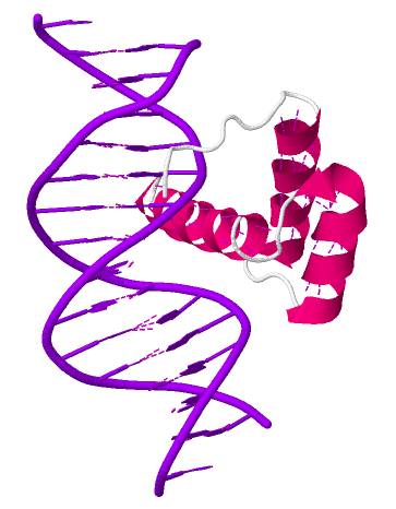
'''General structure of PDX-1 Homeodomain''' | '''General structure of PDX-1 Homeodomain''' | ||
| + | |||
| + | |||
This PDB structure corresponds to the PDX-1 homeodomain. This part of the transcription factor folds into three α-helices (pink color) and a flexible N-terminal arm (white-grey color). This homeodomain interacts with DNA (purple color). | This PDB structure corresponds to the PDX-1 homeodomain. This part of the transcription factor folds into three α-helices (pink color) and a flexible N-terminal arm (white-grey color). This homeodomain interacts with DNA (purple color). | ||
Current revision
General structure of PDX-1 Homeodomain
This PDB structure corresponds to the PDX-1 homeodomain. This part of the transcription factor folds into three α-helices (pink color) and a flexible N-terminal arm (white-grey color). This homeodomain interacts with DNA (purple color).
References : http://www.rcsb.org/pdb/explore/jmol.do?structureId=2H1K&bionumber=1
File history
Click on a date/time to view the file as it appeared at that time.
| Date/Time | User | Dimensions | File size | Comment | |
|---|---|---|---|---|---|
| (current) | 10:11, 31 December 2013 | Megane Denu (Talk | contribs) | 363×466 | 24 KB |
- Edit this file using an external application
See the setup instructions for more information.
Links
The following pages link to this file:
