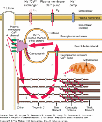
Calsequestrin-2 (or CASQ2) is the soluble Ca2+ binding protein in the sarcoplasmic reticulum lumen of the cardiac muscle cells. CASQ2 could be either in a monomeric, homodimeric, or homooligomeric chain form depending on its bounds with Ca2+. Mutations of CASQ2 are involved in cardiac diseases such as Catecholaminergic Polymorphic Ventricular Tachycardia.

Calsequestrin in the calcium cycle of myocyte contraction
Biological role
The contractions of cardiac myocytes are triggered by the increase of calcium concentration in the cytosol. This phenomenon is highly controlled at several levels. First the calcium is stocked in a cell compartment called the sarcoplasmic reticulum. Then the release of calcium in the cytosol is dependent of the myocytes membrane depolarization. Finally the release of calcium is extremely brief, as soon as the depolarization is over, the calcium is actively pumped in the sarcoplasmic reticulum.
The calsequestrin 2 plays a major role here, because it helps the release of the calcium in the cytosol while the membrane depolarization occurs and traps the calcium inside the lumen of the sarcoplasmic reticulum.
It is also good to notice that a huge release of calcium in the cytosol would be lethal to the cell, since the calcium would precipitate with the free phosphate groups.
Structure
Monomere Structure
Each monomer is divided in : , and the domains. Each of these has a regular structure: a surrounded by .
Usually these domains are involved in redox phenomena, which lead to disulfide bounds creation. Here these domains are inactive but play an important role in the polymerization of CASQ2.
Polymer Structure
Inside the sarcoplasmic reticulum lumen, CASQ2 polymerizes to form , homotetramers and homooligomers.
There are two types of dimerisation: the front-to-front form and the back-to-back form.
The front-to-front form is stabilized by intermolecular interactions between the
of each CASQ2. The intermolecular salt bridges are built between . This dimerisation induces the formation of an electronegative pocket which involves these amino acids: for the first CASQ2 Glu 39, Glu 54, Glu 78, Glu 92, Asp 93 and Asp 101 and for the second CASQ2 Glu 199, Asp 245, Asp 278, Glu 350 and Glu 348.
The back-to-back form is stabilized by intermolecular interactions between the and the
(). The intermolecular salt bridges are built between Glu 215 and Lys 86, Glu 216 and Lys 24, Glu 169 and Lys 85. There is also a hydrogen bond between Ala 82 and Asn 22. This dimerisation induces a very electronegative pocket at the C-terminal region which enables the binding of Ca2+.
Calcium Binding
Each monomere of CASQ2 can bind between 18 to 50 Ca2+. The Ca2+ ions bind to two or more acidic amino acids like or . These amino acids are mainly outside the CASQ2 or in the C-terminal region. It had been shown that Ca2+ binds to an Asp-rich region on the C-terminal domain. When CASQ2 form homooligomers, Ca2+ can bind in the electronegative pocket due to the front-to-front form and back-to-back form.
Ca2+ is not the only ion which can bind to the CASQ2. One of them is Mg2+. The affinity is for Mg2+ is lower than the affinity for Ca2+ however the number of Ca2+ decrease. Another ion is H+. When the pH is low, more H+ will bind to the acidic amino acids and they can not bind Ca2+ anymore.
Interaction between CASQ2 and RYR
Binding sites
CASQ2 is anchored into the membrane of SR thanks to two integral proteins: the triadin and the junctin. Triadin as well as Juctin can bind to CASQ2 because of its KEKE motif between the amino acids 210 and 224 for the triadin. The binding site of CASQ2 for the both protein is the Asp-rich region of the C-terminal region.
Triadin and Junctin interact with Ryanodin Receptor (RyR).
The binding site of CASQ2 to RyR is unknow.
Consequences of the bound of CASQ2
When CASQ2 binds to Triadin and Junctin, it induces the inhibition of RyR and when CASQ2 unbinds Triadin and Junctin , it induces the activation of Ryr and an efflux of Ca2+ from the SR to the cytoplasm. CASQ2 is free when the concentration of Ca2+ is higher than 1 mM in the SR lumen.
Regulation of CASQ2
CASQ2 can be phosphorylated by three different kinases: casein kinase I (CK I), casein kianse II (CK II) and ε protein kinase C1 (εPKC1). CK II is located in the SR and is able to phosphorylate Ser 378, Ser 382 and Ser 386. These residues are on the C-terminal domain. The consensus sequence recognized by CK II is Ser/Thr-X-X-Asp/Glu. More there are acidic residues after this consensus sequence, more the probabilty of phosphorylation increases.
"The phosphorylation and de-phosphorylation of CASQ2 my provide an off/on switch for CASQ2 to regulate Ca2+" But there is not any prove yet.
The p
There are and .
can bind the Ca2+ especially the and the .
The Ct domain is highly implicated in the Ca2+ bounds.
The Ca2+ is bound with the interaction of at least 2 acidic amino acids (glutamate or aspartate). These amino acids are in the external part of the protein. When Ca2+ binds to these amino acids there is a structural change which increase the number of alpha helix. Without Ca2+, there are 10-13% of alpha helix but in presence of Ca2+ there 20->35% of alpha helix.
The N-term domain is implicated in front-to-front dimer interactions. While the C-term domain is involved in back-to-back dimer interactions.

