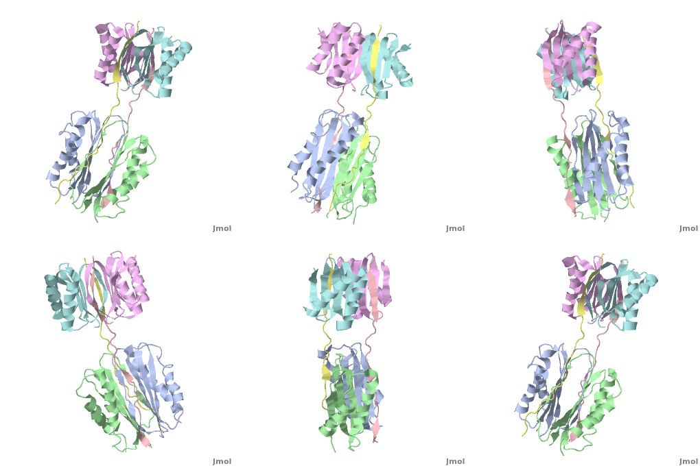
Dynein is a motor protein which walks along the microtubule toward its minus end, i.e. it is a minus-end directed motor. Dyneins are classified as cytoplasmic (DYNC) or axonemal. The Dyneins are composed of heavy, intermediate and light chains. For discussion of the DYNC inter mediate and light chains see: Dynein light and intermediate chain.
Quaternary Structure of Drosophila melanogaster IC/Tctex-1/LC8; Allosteric Interactions of Dynein Light Chains with Dynein Intermediate Chain
Cytoplasmic Dynein is a motorprotein, which plays an important role in many cellular processes as vesicular transport, mitosis , and cell migration.
Dynein consists of four homodimeric subunits named heavychain (HC) (~530kDa), intermediate chain (IC) (74kDa), light intermediate chain (LIC) (two members, 30 and 50kDa), and light chain (LC) (three members, 10, 12, and 14kDa). On the C-terminal region the HCs bind to mircrotubules and they hydrolyse ATP to generate force towards the minus end of the microtubule. On the N-terminal region they self-associate with the LICs and the ICs. Also the LICs and the ICs associate together. And the ICs bind to the LCs as the following article describes. The whole comlpex then binds to cargo.
Earlier research considered that every LC subunit binds cargo like transcription factors, signaling molecules, and scaffolding proteins, and links this cargo to the motor complex. This would allow the retrograde transport of the cargo along microtubules through the cytoplasm. Recent structural and thermodynamic studies do not support this mechanism. Due to the fact that the LC binding IC parts block the major cargo binding sites on the LC. However, in this article we want to focus on the interaction between the LC and the IC.
Structural Analysis
Overall Structure
The overall structure of this part of the Dynein complex consists of two homodimeric LCs named and and two parts of the . The associating sites of the IC strands are β-sheets. Every subunit of every light chain interacts with one β-strand of one part of the IC. This IC β-sheet lies in a on the outside of the LC dimer. The IC strands run roughly parallel to the fold axis of the LCs. In fact the binding sites of LC8 and TcTex1 are not parallel and so the IC strand shows a bend.
Intermediate Chain
The intermediate chain residues LEU110-SER-VAL112 and ASN119-ILE120 () interact with TcTex1 and TYR128-THR-LYS-GLN-THR-GLN133 () interact wit LC8. Each associating part undergoes a disorder-to-order transition to form β-strands incorporating the β-sheets in the LC.
TcTex1
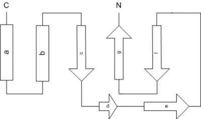
The structure of TcTex1 consists, as shown in the band diagram, of two α-helices ((a) and (b)) and five β-sheets ((c)-(g)). The α-helices are found exposed to the outside of the globular subunit and so they consist of charged and polar residues on the solvent exposed side whereas the buried residues are mostly aliphatic. Also every second residue of the solvent exposed β-sheet (f) is polar and the others apolar.
The binding sites of TcTex1 are the β-sheets (d) and (e). As said before they lie in a cleft on the outside and due to that the IC (or cargo) binds with hydrophobic residues to it.
LC8
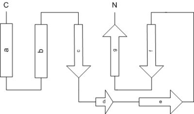
LC8 consists of two α-helices and four β-sheets as shown in the band diagram. We find analogical to TcTex1 the globular assembly. And as in TcTex1 the α-helices are found on the solvent exposed side with polar residues to the outside and aliphatic residues to the center of the subunit. β-sheet (a) is also solvent exposed but unlike the solvent exposed β-sheet in TcTex1 the residues looking into the solvent are not charged. Contrariwise, they are aliphatic.
Discussion
Latest structural research on Dynein has brought a different view on the behaviour of the Dynein-LC and its association with the IC. It was thought that the LCs bind cargo to the Dynein complex. Due to the fact that the IC binds at the same cleft as cargo would, there is a conflict between the two binding partners and we have to consider a new scenario for the interaction between LCs and the motor complex.
Dynein transports different cargo and to have influence on this transports could be interesting as it could be the case in stopping the transport of viral products. But as we see our previous knowledge is limited and so further research on this multisubunit protein is preferable.
3D Structures of Dynein
Updated on 27-February-2014
Dynein light chain
3dvt, 3bri - DmDYNC light chain - Drosophila melanogaster
3dvh - DmDYNC light chain (mutant)
1rhw - DmDYNC light chain - NMR
1ygt - DmDYNC light chain TcTex1
2e8j, 2b95, 1z09 - hDYNC light chain 2A – human – NMR
2hz5 - hDYNC light chain 2A
2xqq - hDYNC light chain 2 + peptide
3p8m - hDYNC light chain 2 + GCN4
3zke, 3zkf - hDYNC light chain + NEK9 peptide
3tq7 – hDYNC + EB1C/EB3C
1y4o - mDYNC light chain 2A – mouse
1re6 - mDYNC light chain 2 – NMR
1pwj, 1pwk, 1f3c - rDYNC light chain 2 – rat – NMR
1f95, 1f96 - rDYNC light chain + peptide – NMR
3dvp, 3brl, 3e2b - DmDYNC light chain + peptide
1yo3 – DYNC light chain 1 – Plasmodium falciparum
1xdx, 1m9l, 1ds9 - CrDYNC light chain TcTex1 – Chlamydomonas reinhardtii – NMR
4ds1 - yDYNC light chain + nucleoporin Nup129 peptide - yeast
4ht6 – yDYNC + PAC11 peptide
3rjs - DYNC light chain – Toxoplasma gondii
Dynein intermediate chain
3l7h – DmDYNC intermediate chain light chain roadblock
3l9k - DmDYNC intermediate chain light chain roadblock + intermediate chain
3fm7, 3glw, 2pg1, 2p2t - DmDYNC intermediate chain + light chain
Dynein heavy chain
3qmz – yDYNC heavy chain + glutathione-S-transferase
3err - mDYNC heavy chain microtubule-binding domain/Ser-tRNA synthetase
3j1t - mDYNC heavy chain + tubulin α-1B chain
3j1u - mDYNC heavy chain + tubulin α-1B+β-2B chains
3ay1, 3vkg, 3vkh - DYNC heavy chain – Dictyostelium discoideum
2rr7 – CrDYNC microtubule-binding domain
4aki, 4akg – yDYNC motor domain + ATP
4akh – yDYNC motor domain + AMPPNP
4ai6 – yDYNC motor domain + ADP<br /


