We apologize for Proteopedia being slow to respond. For the past two years, a new implementation of Proteopedia has been being built. Soon, it will replace this 18-year old system. All existing content will be moved to the new system at a date that will be announced here.
User:Daud Akhtar/Sandbox 1
From Proteopedia
(Difference between revisions)
| Line 10: | Line 10: | ||
Glucose 6-Phosphate Dehydrogenase (G6PD) is a metabolic X-linked enzyme, which catalyzes the conversion of glucose-6-phosphate (G6P) to 6-phosphoglucono-δ-lactone<ref>PMID: 11375432 </ref> . This redox reaction is the first and rate-determining step in the pentose phosphate pathway in which coenzyme NADP+ is reduced through the transfer of a hydride from G6P . Consequently, NADPH is generated and helps restore reduced glutathione (GSH), which is an important anti-oxidant. NADPH and GSH thereby helping protect cells from oxidative stress by converting peroxides into water. (2). G6PD is most abundant in intracellular fluid and is conserved over a large array of different organisms. Specifically, higher plants exhibit several isoforms of G6PD in different cellular locations such as the cytosol, the plastidic stroma, and peroxisomes. [2] In humans, active G6PD generally exists as a dimer/tetramer equilibrium, which is depending on pH and ionic strength. At high pH and ionic strength, the equilibrium is shifted towards the dimer, whereas low pH conditions cause a shift to the tetramer. Each monomer of G6PD consists of 514 amino acids with a molecular weight of 59 kDa, More than 400 different variants of G6PD have been identified where these variants differ in the location of point mutations in the G6PD gene. The G6PD gene is located on the chromosome Xq28 region. These mutation results in deficiencies, which range from mild to severe phenotypic abnormalities such as hemolytic anemia. The exposure of erythrocytes to oxidative stress due to a lack of NADPH in cells results in their destruction causing hemolytic anemia. | Glucose 6-Phosphate Dehydrogenase (G6PD) is a metabolic X-linked enzyme, which catalyzes the conversion of glucose-6-phosphate (G6P) to 6-phosphoglucono-δ-lactone<ref>PMID: 11375432 </ref> . This redox reaction is the first and rate-determining step in the pentose phosphate pathway in which coenzyme NADP+ is reduced through the transfer of a hydride from G6P . Consequently, NADPH is generated and helps restore reduced glutathione (GSH), which is an important anti-oxidant. NADPH and GSH thereby helping protect cells from oxidative stress by converting peroxides into water. (2). G6PD is most abundant in intracellular fluid and is conserved over a large array of different organisms. Specifically, higher plants exhibit several isoforms of G6PD in different cellular locations such as the cytosol, the plastidic stroma, and peroxisomes. [2] In humans, active G6PD generally exists as a dimer/tetramer equilibrium, which is depending on pH and ionic strength. At high pH and ionic strength, the equilibrium is shifted towards the dimer, whereas low pH conditions cause a shift to the tetramer. Each monomer of G6PD consists of 514 amino acids with a molecular weight of 59 kDa, More than 400 different variants of G6PD have been identified where these variants differ in the location of point mutations in the G6PD gene. The G6PD gene is located on the chromosome Xq28 region. These mutation results in deficiencies, which range from mild to severe phenotypic abnormalities such as hemolytic anemia. The exposure of erythrocytes to oxidative stress due to a lack of NADPH in cells results in their destruction causing hemolytic anemia. | ||
Glucose 6 Phophate Dehydrogenease is an enzyme that plays a key role in the pentose phosphate pathway <ref>PMID: 11375432 </ref> | Glucose 6 Phophate Dehydrogenease is an enzyme that plays a key role in the pentose phosphate pathway <ref>PMID: 11375432 </ref> | ||
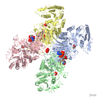
| - | <scene name='58/580852/Ligand/4'>Ligands on each respective subunit of a G6PD tetramer(ABCD) where ligand color corresponds to the respective subunit(A=blue,B=green,C=pink,D=yellow) </scene> | + | <scene name='58/580852/Ligand/4'>Ligands(both G6P and coenzyme NADP+) on each respective subunit of a G6PD tetramer(ABCD) where ligand color corresponds to the respective subunit(A=blue,B=green,C=pink,D=yellow) </scene> |
==Species Distribution== | ==Species Distribution== | ||
| Line 19: | Line 19: | ||
===NADP+ Binding Domain=== | ===NADP+ Binding Domain=== | ||
| - | The first domain is a NADP+ binding domain with a β-β fold. The binding site for the coenzyme NADP+ is between the first β-sheet and the α-helix with a finger print sequence of GASGDLA (residues 38-44). At the C-terminus of the β-sheet, a highly conserved Arginine (residue 72) functions to bind the 2’phosphate of NADP to increase specificity. | + | The first domain is a <scene name='58/580852/Nadp_binding_site/1'>NADP+ binding domain</scene> with a β-β fold. The binding site for the coenzyme NADP+ is between the first β-sheet and the α-helix with a finger print sequence of GASGDLA (residues 38-44). At the C-terminus of the β-sheet, a highly conserved Arginine (residue 72) functions to bind the 2’phosphate of NADP to increase specificity. |
===Substrate(Glucose-6-Phosphate) Binding Domain=== | ===Substrate(Glucose-6-Phosphate) Binding Domain=== | ||
Revision as of 00:46, 30 March 2014
Glucose-6-Phosphate Dehydrogenase(G6PD)
| |||||||||||
Glucose 6 Phosphate Dehydrognease
jghgjgjhgjhg
References
- ↑ Salati LM, Amir-Ahmady B. Dietary regulation of expression of glucose-6-phosphate dehydrogenase. Annu Rev Nutr. 2001;21:121-40. PMID:11375432 doi:http://dx.doi.org/10.1146/annurev.nutr.21.1.121
- ↑ Salati LM, Amir-Ahmady B. Dietary regulation of expression of glucose-6-phosphate dehydrogenase. Annu Rev Nutr. 2001;21:121-40. PMID:11375432 doi:http://dx.doi.org/10.1146/annurev.nutr.21.1.121
- ↑ . Glucose-6-phosphate dehydrogenase deficiency. WHO Working Group. Bull World Health Organ. 1989;67(6):601-11. PMID:2633878
- ↑ Au SW, Gover S, Lam VM, Adams MJ. Human glucose-6-phosphate dehydrogenase: the crystal structure reveals a structural NADP(+) molecule and provides insights into enzyme deficiency. Structure. 2000 Mar 15;8(3):293-303. PMID:10745013
- ↑ Manganelli G, Masullo U, Passarelli S, Filosa S. Glucose-6-phosphate dehydrogenase deficiency: disadvantages and possible benefits. Cardiovasc Hematol Disord Drug Targets. 2013 Mar 1;13(1):73-82. PMID:23534950
- ↑ Beutler E. Glucose-6-phosphate dehydrogenase deficiency. N Engl J Med. 1991 Jan 17;324(3):169-74. PMID:1984194 doi:http://dx.doi.org/10.1056/NEJM199101173240306
