We apologize for Proteopedia being slow to respond. For the past two years, a new implementation of Proteopedia has been being built. Soon, it will replace this 18-year old system. All existing content will be moved to the new system at a date that will be announced here.
JMS/sandbox15
From Proteopedia
(Difference between revisions)
| Line 19: | Line 19: | ||
The ability of an increase in number of positively charged amino acids and/or decrease in the number of negatively charged amino acids to enable higher solubility is a known phenomenon<ref>doi: 10.1073/pnas.0402797101</ref>, and this study is consistent with previous reports<ref>PMID: 14741208</ref>. The aquatic animals have increased their net charge in a variety of ways - different combinations of amino acids switches. We present one such manifestation of this overall trend, by comparing the elephant and whale myoglobin structures. | The ability of an increase in number of positively charged amino acids and/or decrease in the number of negatively charged amino acids to enable higher solubility is a known phenomenon<ref>doi: 10.1073/pnas.0402797101</ref>, and this study is consistent with previous reports<ref>PMID: 14741208</ref>. The aquatic animals have increased their net charge in a variety of ways - different combinations of amino acids switches. We present one such manifestation of this overall trend, by comparing the elephant and whale myoglobin structures. | ||
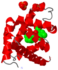
| - | Out of 27 divergent amino acids between whale and elephant's myoglobin - from a total of 153 amino acids - only 8 of these amino acids lead to changes of charge. These eight amino acids are shown for whales and elephants, <scene name='57/575026/Electrostatics/19'>side by side</scene>. Arginine and Lysine have a charge of +1, aspartic and glutamic acids have charges of -1, and histidine in positions 12 and 116 have a charge of about +0.5 (supplementary Table S2 <ref name="whaleMyo" />). The whale amino acids, have an illustrative eletrostatic field drawn around the <scene name='57/575026/Electrostatics/17'>electrically charged atom</scene> in the residue. Next to the whale amino acid, the <scene name='57/575026/Electrostatics/18'>elephant residues</scene> are shown in yellow halos and labelled with the residue name. From studying the differences between these two proteins, it is clear that the whale protein has more areas with a positive electrostatic field. These positive electrostatic fields are <scene name='57/575026/Electrostatics/19'>scattered about the surface</scene> of the whale protein, and will repel any whale myoglobin, preventing the protein-protein interactions that lead to aggregation. | + | Out of 27 divergent amino acids between whale and elephant's myoglobin - from a total of 153 amino acids - only 8 of these amino acids lead to changes of charge. These eight amino acids are shown for whales and elephants, <scene name='57/575026/Electrostatics/19'>side by side</scene>. Arginine and Lysine have a charge of +1, aspartic and glutamic acids have charges of -1, and histidine in positions 12 and 116 have a charge of about +0.5 (supplementary Table S2 <ref name="whaleMyo" />). The whale amino acids, have an illustrative eletrostatic field drawn around the <scene name='57/575026/Electrostatics/17'>electrically charged atom</scene> in the residue. Note that the effective size of the electric field of the charged atom at the end of residues like lysine is actually larger, because of the multiple conformations a long residue <scene name='57/575026/Electrostatics/17'>like lysine</scene> moves between. Next to the whale amino acid, the <scene name='57/575026/Electrostatics/18'>elephant residues</scene> are shown in yellow halos and labelled with the residue name. From studying the differences between these two proteins, it is clear that the whale protein has more areas with a positive electrostatic field. These positive electrostatic fields are <scene name='57/575026/Electrostatics/19'>scattered about the surface</scene> of the whale protein, and will repel any whale myoglobin, preventing the protein-protein interactions that lead to aggregation. |
| - | Another way to say this, in the case of proteins <scene name='57/575026/Electrostatics/16'>in solvents such as water</scene>, is that, for each and every protein, <scene name='57/575026/Electrostatics/21'>water binds to the hydrogens</scene> (e.g., lysine's ammonium at physiology pH has three hydrogens - not shown) coming off a positively charged atom, or associates with the charged atom itself, thus screening each protein from the other proteins | + | Another way to say this, in the case of proteins <scene name='57/575026/Electrostatics/16'>in solvents such as water</scene>, is that, for each and every protein, <scene name='57/575026/Electrostatics/21'>water binds to the hydrogens</scene> (e.g., lysine's ammonium at physiology pH has three hydrogens - not shown) coming off a positively charged atom, or associates with the charged atom itself, thus screening each protein from the other proteins. |
| - | This picture, while illustrative of the basic principles of how small electrostatic fields changes can greatly increase a protein's solubility, is incomplete. | + | This picture, while illustrative of the basic principles of how small electrostatic fields changes can greatly increase a protein's solubility, is incomplete. Solvated ions, crowding conditions, and neighboring residues to these divergent residues, all affect the electrostatic field. For example, it may be in some cases, that a negatively charged residue neigbors one of these additional positive residues, and from a distance relatively larger than the distance between the two oppositely charged residues, the electrostatic field is zero. |
<!-- | <!-- | ||
Revision as of 17:48, 28 April 2014
| |||||||||||
References:
- ↑ 1.0 1.1 Mirceta S, Signore AV, Burns JM, Cossins AR, Campbell KL, Berenbrink M. Evolution of mammalian diving capacity traced by myoglobin net surface charge. Science. 2013 Jun 14;340(6138):1234192. doi: 10.1126/science.1234192. PMID:23766330 doi:http://dx.doi.org/10.1126/science.1234192
- ↑ Brocchieri L. Environmental signatures in proteome properties. Proc Natl Acad Sci U S A. 2004 Jun 1;101(22):8257-8. Epub 2004 May 24. PMID:15159533 doi:http://dx.doi.org/10.1073/pnas.0402797101
- ↑ Goh CS, Lan N, Douglas SM, Wu B, Echols N, Smith A, Milburn D, Montelione GT, Zhao H, Gerstein M. Mining the structural genomics pipeline: identification of protein properties that affect high-throughput experimental analysis. J Mol Biol. 2004 Feb 6;336(1):115-30. PMID:14741208 doi:http://dx.doi.org/10.1016/S0022283603014748

