This old version of Proteopedia is provided for student assignments while the new version is undergoing repairs. Content and edits done in this old version of Proteopedia after March 1, 2026 will eventually be lost when it is retired in about June of 2026.
Apply for new accounts at the new Proteopedia. Your logins will work in both the old and new versions.
Image:Thioredoxin fold.jpg
From Proteopedia
Size of this preview: 452 × 599 pixels
Full resolution (531 × 704 pixel, file size: 36 KB, MIME type: image/jpeg)
Drew Barber (Talk | contribs)
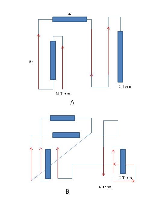
(Figure 4. Thioredoxin Fold. Red arrows represent B-sheets while blue rectangles represent alpha-helices. A. Thioredoxin fold. B. Fold of glutathione peroxidase.)
Next diff →
Current revision
Figure 4. Thioredoxin Fold. Red arrows represent B-sheets while blue rectangles represent alpha-helices. A. Thioredoxin fold. B. Fold of glutathione peroxidase.
File history
Click on a date/time to view the file as it appeared at that time.
| Date/Time | User | Dimensions | File size | Comment | |
|---|---|---|---|---|---|
| (current) | 07:31, 30 April 2014 | Drew Barber (Talk | contribs) | 531×704 | 36 KB | Figure 4. Thioredoxin Fold. Red arrows represent B-sheets while blue rectangles represent alpha-helices. A. Thioredoxin fold. B. Fold of glutathione peroxidase. |
- Edit this file using an external application
See the setup instructions for more information.
Links
The following pages link to this file:
Metadata
This file contains additional information, probably added from the digital camera or scanner used to create or digitize it. If the file has been modified from its original state, some details may not fully reflect the modified image.
| Orientation | Normal |
|---|
