We apologize for Proteopedia being slow to respond. For the past two years, a new implementation of Proteopedia has been being built. Soon, it will replace this 18-year old system. All existing content will be moved to the new system at a date that will be announced here.
3tj8
From Proteopedia
(Difference between revisions)
| Line 1: | Line 1: | ||
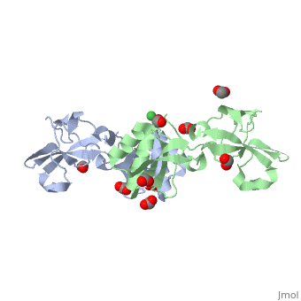
| - | [[ | + | ==Crystal structure of Helicobacter pylori UreE bound to Ni2+== |
| + | <StructureSection load='3tj8' size='340' side='right' caption='[[3tj8]], [[Resolution|resolution]] 1.59Å' scene=''> | ||
| + | == Structural highlights == | ||
| + | <table><tr><td colspan='2'>[[3tj8]] is a 2 chain structure with sequence from [http://en.wikipedia.org/wiki/Helicobacter_pylori Helicobacter pylori]. Full crystallographic information is available from [http://oca.weizmann.ac.il/oca-bin/ocashort?id=3TJ8 OCA]. For a <b>guided tour on the structure components</b> use [http://oca.weizmann.ac.il/oca-docs/fgij/fg.htm?mol=3TJ8 FirstGlance]. <br> | ||
| + | </td></tr><tr><td class="sblockLbl"><b>[[Ligand|Ligands:]]</b></td><td class="sblockDat"><scene name='pdbligand=FMT:FORMIC+ACID'>FMT</scene>, <scene name='pdbligand=NI:NICKEL+(II)+ION'>NI</scene><br> | ||
| + | <tr><td class="sblockLbl"><b>[[Related_structure|Related:]]</b></td><td class="sblockDat">[[3tj9|3tj9]], [[3tja|3tja]]</td></tr> | ||
| + | <tr><td class="sblockLbl"><b>[[Gene|Gene:]]</b></td><td class="sblockDat">ureE, HP_0070 ([http://www.ncbi.nlm.nih.gov/Taxonomy/Browser/wwwtax.cgi?mode=Info&srchmode=5&id=210 Helicobacter pylori])</td></tr> | ||
| + | <tr><td class="sblockLbl"><b>Resources:</b></td><td class="sblockDat"><span class='plainlinks'>[http://oca.weizmann.ac.il/oca-docs/fgij/fg.htm?mol=3tj8 FirstGlance], [http://oca.weizmann.ac.il/oca-bin/ocaids?id=3tj8 OCA], [http://www.rcsb.org/pdb/explore.do?structureId=3tj8 RCSB], [http://www.ebi.ac.uk/pdbsum/3tj8 PDBsum]</span></td></tr> | ||
| + | <table> | ||
| + | <div style="background-color:#fffaf0;"> | ||
| + | == Publication Abstract from PubMed == | ||
| + | The survival and growth of the pathogen Helicobacter pylori in the gastric acidic environment is ensured by the activity of urease, an enzyme containing two essential Ni2+ ions in the active site. The metallo-chaperone UreE facilitates in vivo Ni2+ insertion into the apo-enzyme. Crystals of apo-HpUreE and its Ni2+ and Zn2+ bound forms were obtained from protein solutions in the absence and presence of the metal ions. The crystal structures of the homodimeric protein, determined at 2.00 A (apo), 1.59 A (Ni) and 2.52 A (Zn) resolution, show the conserved proximal and solvent-exposed His102 residues from two adjacent monomers invariably involved in metal binding. The C-terminal regions of the apo-protein are disordered in the crystal, but acquire significant ordering in the presence of the metal ions due to the binding of His152. The analysis of X-ray absorption spectral data obtained on solutions of Ni2+- and Zn2+-HpUreE provided accurate information of the metal ion environment in the absence of solid-state effects. These results reveal the role of the histidine residues at the protein C-terminus in metal ion binding, and the mutual influence of protein framework and metal ion stereo-electronic properties in establishing coordination number and geometry leading to metal selectivity. | ||
| - | + | Crystallographic and X-ray absorption spectroscopic characterization of Helicobacter pylori UreE bound to Ni2+ and Zn2+ reveal a role for the disordered C-terminal arm in metal trafficking.,Banaszak K, Martin-Diaconescu V, Bellucci M, Zambelli B, Rypniewski W, Maroney MJ, Ciurli S Biochem J. 2011 Oct 20. PMID:22010876<ref>PMID:22010876</ref> | |
| - | + | ||
| - | + | ||
| - | + | ||
| - | + | ||
| - | + | ||
| - | + | ||
| - | + | From MEDLINE®/PubMed®, a database of the U.S. National Library of Medicine.<br> | |
| - | + | </div> | |
| - | + | == References == | |
| - | + | <references/> | |
| - | + | __TOC__ | |
| - | + | </StructureSection> | |
| - | + | ||
| - | + | ||
| - | + | ||
| - | + | ||
| - | + | ||
| - | + | ||
| - | == | + | |
| - | < | + | |
[[Category: Helicobacter pylori]] | [[Category: Helicobacter pylori]] | ||
[[Category: Banaszak, K.]] | [[Category: Banaszak, K.]] | ||
Revision as of 05:58, 5 June 2014
Crystal structure of Helicobacter pylori UreE bound to Ni2+
| |||||||||||