This old version of Proteopedia is provided for student assignments while the new version is undergoing repairs. Content and edits done in this old version of Proteopedia after March 1, 2026 will eventually be lost when it is retired in about June of 2026.
Apply for new accounts at the new Proteopedia. Your logins will work in both the old and new versions.
Catalase 2CAG bcce2014
From Proteopedia
(Difference between revisions)
| Line 1: | Line 1: | ||
---- | ---- | ||
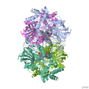
| - | == | + | == Catalase file created from PDB 2CAG at BCCE2014 workshop == |
<StructureSection load='2cag' size='340' side='right' caption='Caption for this structure' scene=''> | <StructureSection load='2cag' size='340' side='right' caption='Caption for this structure' scene=''> | ||
This is a default text for your page '''Sandbox bcce36'''. Click above on '''edit this page''' to modify. Be careful with the < and > signs. | This is a default text for your page '''Sandbox bcce36'''. Click above on '''edit this page''' to modify. Be careful with the < and > signs. | ||
| Line 6: | Line 6: | ||
== Function == | == Function == | ||
| - | + | This catalase protects Red blood cells from Reactive Oxygen Species (ROS) such as H2O2. | |
== Structural highlights == | == Structural highlights == | ||
| - | This is catalase 2cag <scene name='59/596494/Initial/1'>Overall | + | This is catalase 2cag <scene name='59/596494/Initial/1'>Overall struacture</scene> |
Zooming in on the <scene name='59/596494/Active_site/1'>active site</scene>. | Zooming in on the <scene name='59/596494/Active_site/1'>active site</scene>. | ||
| - | The | + | The <scene name='59/596494/Proximal_ligand_binding_site/1'>proximal ligand</scene> is Tyrosine337. Distal residues His54 and Asn127 are also shown. |
This is a sample scene created with SAT to <scene name="/12/3456/Sample/1">color</scene> by Group, and another to make <scene name="/12/3456/Sample/2">a transparent representation</scene> of the protein. You can make your own scenes on SAT starting from scratch or loading and editing one of these sample scenes. | This is a sample scene created with SAT to <scene name="/12/3456/Sample/1">color</scene> by Group, and another to make <scene name="/12/3456/Sample/2">a transparent representation</scene> of the protein. You can make your own scenes on SAT starting from scratch or loading and editing one of these sample scenes. | ||
Revision as of 03:53, 7 August 2014
Catalase file created from PDB 2CAG at BCCE2014 workshop
| |||||||||||
References
- ↑ Hanson, R. M., Prilusky, J., Renjian, Z., Nakane, T. and Sussman, J. L. (2013), JSmol and the Next-Generation Web-Based Representation of 3D Molecular Structure as Applied to Proteopedia. Isr. J. Chem., 53:207-216. doi:http://dx.doi.org/10.1002/ijch.201300024
- ↑ Herraez A. Biomolecules in the computer: Jmol to the rescue. Biochem Mol Biol Educ. 2006 Jul;34(4):255-61. doi: 10.1002/bmb.2006.494034042644. PMID:21638687 doi:10.1002/bmb.2006.494034042644
