This old version of Proteopedia is provided for student assignments while the new version is undergoing repairs. Content and edits done in this old version of Proteopedia after March 1, 2026 will eventually be lost when it is retired in about June of 2026.
Apply for new accounts at the new Proteopedia. Your logins will work in both the old and new versions.
2wwp
From Proteopedia
| Line 1: | Line 1: | ||
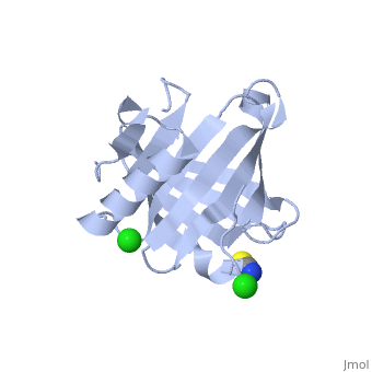
| - | [[ | + | ==Crystal structure of the human lipocalin-type prostaglandin D synthase== |
| + | <StructureSection load='2wwp' size='340' side='right' caption='[[2wwp]], [[Resolution|resolution]] 2.00Å' scene=''> | ||
| + | == Structural highlights == | ||
| + | <table><tr><td colspan='2'>[[2wwp]] is a 2 chain structure with sequence from [http://en.wikipedia.org/wiki/Human Human]. Full crystallographic information is available from [http://oca.weizmann.ac.il/oca-bin/ocashort?id=2WWP OCA]. For a <b>guided tour on the structure components</b> use [http://oca.weizmann.ac.il/oca-docs/fgij/fg.htm?mol=2WWP FirstGlance]. <br> | ||
| + | </td></tr><tr><td class="sblockLbl"><b>[[Ligand|Ligands:]]</b></td><td class="sblockDat"><scene name='pdbligand=CL:CHLORIDE+ION'>CL</scene>, <scene name='pdbligand=SCN:THIOCYANATE+ION'>SCN</scene><br> | ||
| + | <tr><td class="sblockLbl"><b>Activity:</b></td><td class="sblockDat"><span class='plainlinks'>[http://en.wikipedia.org/wiki/Prostaglandin-D_synthase Prostaglandin-D synthase], with EC number [http://www.brenda-enzymes.info/php/result_flat.php4?ecno=5.3.99.2 5.3.99.2] </span></td></tr> | ||
| + | <tr><td class="sblockLbl"><b>Resources:</b></td><td class="sblockDat"><span class='plainlinks'>[http://oca.weizmann.ac.il/oca-docs/fgij/fg.htm?mol=2wwp FirstGlance], [http://oca.weizmann.ac.il/oca-bin/ocaids?id=2wwp OCA], [http://www.rcsb.org/pdb/explore.do?structureId=2wwp RCSB], [http://www.ebi.ac.uk/pdbsum/2wwp PDBsum]</span></td></tr> | ||
| + | <table> | ||
| + | == Evolutionary Conservation == | ||
| + | [[Image:Consurf_key_small.gif|200px|right]] | ||
| + | Check<jmol> | ||
| + | <jmolCheckbox> | ||
| + | <scriptWhenChecked>select protein; define ~consurf_to_do selected; consurf_initial_scene = true; script "/wiki/ConSurf/ww/2wwp_consurf.spt"</scriptWhenChecked> | ||
| + | <scriptWhenUnchecked>script /wiki/extensions/Proteopedia/spt/initialview01.spt</scriptWhenUnchecked> | ||
| + | <text>to colour the structure by Evolutionary Conservation</text> | ||
| + | </jmolCheckbox> | ||
| + | </jmol>, as determined by [http://consurfdb.tau.ac.il/ ConSurfDB]. You may read the [[Conservation%2C_Evolutionary|explanation]] of the method and the full data available from [http://bental.tau.ac.il/new_ConSurfDB/chain_selection.php?pdb_ID=2ata ConSurf]. | ||
| + | <div style="clear:both"></div> | ||
| + | <div style="background-color:#fffaf0;"> | ||
| + | == Publication Abstract from PubMed == | ||
| + | Lipocalin prostaglandin D synthase (L-PGDS) regulates synthesis of an important inflammatory and signaling mediator, prostaglandin D2 (PGD2). Here, we used structural, biophysical, and biochemical approaches to address the mechanistic aspects of substrate entry, catalysis, and product exit of this enzyme. Structure of human L-PGDS was solved in a complex with a substrate analog (SA) and in ligand-free form. Its catalytic Cys 65 thiol group was found in two different conformations, each making a distinct hydrogen bond network to neighboring residues. These help in elucidating the mechanism of the cysteine nucleophile activation. Electron density for ligand observed in the active site defined the substrate binding regions, but did not allow unambiguous fitting of the SA. To further understand ligand binding, we used NMR spectroscopy to map the binding sites and to show the dynamics of protein-substrate and protein-product interactions. A model for ligand binding at the catalytic site is proposed, showing a second binding site involved in ligand exit and entry. NMR chemical shift perturbations and NMR resonance line-width alterations (observed as changes of intensity in two-dimensional cross-peaks in [(1)H,(15)N]-transfer relaxation optimization spectroscopy) for residues at the Omega loop (A-B loop), E-F loop, and G-H loop besides the catalytic sites indicate involvement of these residues in ligand entry/egress. | ||
| - | + | Structural and dynamic insights into substrate binding and catalysis of human lipocalin prostaglandin D synthase.,Lim SM, Chen D, Teo H, Roos A, Jansson AE, Nyman T, Tresaugues L, Pervushin K, Nordlund P J Lipid Res. 2013 Jun;54(6):1630-43. doi: 10.1194/jlr.M035410. Epub 2013 Mar 22. PMID:23526831<ref>PMID:23526831</ref> | |
| - | + | From MEDLINE®/PubMed®, a database of the U.S. National Library of Medicine.<br> | |
| - | + | </div> | |
| - | + | ||
| - | + | ||
| - | + | ||
==See Also== | ==See Also== | ||
*[[Prostaglandin D synthase|Prostaglandin D synthase]] | *[[Prostaglandin D synthase|Prostaglandin D synthase]] | ||
| - | [[Category: | + | == References == |
| + | <references/> | ||
| + | __TOC__ | ||
| + | </StructureSection> | ||
| + | [[Category: Human]] | ||
[[Category: Prostaglandin-D synthase]] | [[Category: Prostaglandin-D synthase]] | ||
[[Category: Arrowsmith, C H.]] | [[Category: Arrowsmith, C H.]] | ||
| Line 33: | Line 54: | ||
[[Category: Moche, M.]] | [[Category: Moche, M.]] | ||
[[Category: Nielsen, T K.]] | [[Category: Nielsen, T K.]] | ||
| - | [[Category: Nordlund, P.]] | ||
[[Category: Nyman, T.]] | [[Category: Nyman, T.]] | ||
[[Category: Persson, C.]] | [[Category: Persson, C.]] | ||
| Line 39: | Line 59: | ||
[[Category: Schuler, H.]] | [[Category: Schuler, H.]] | ||
[[Category: Schutz, P.]] | [[Category: Schutz, P.]] | ||
| - | [[Category: Sgc, Structural Genomics Consortium.]] | + | [[Category: Sgc, P Nordlund Structural Genomics Consortium.]] |
[[Category: Siponen, M I.]] | [[Category: Siponen, M I.]] | ||
[[Category: Svensson, L.]] | [[Category: Svensson, L.]] | ||
Revision as of 06:44, 13 August 2014
Crystal structure of the human lipocalin-type prostaglandin D synthase
| |||||||||||

Categories: Human | Prostaglandin-D synthase | Arrowsmith, C H. | Berg, S Van Den. | Berglund, H. | Bountra, C. | Collins, R. | Edwards, A M. | Flodin, S. | Flores, A. | Graslund, S. | Hammarstrom, M. | Johansson, A. | Johansson, I. | Kallas, A. | Karlberg, T. | Kotyenova, T. | Kotzch, A. | Kraulis, P. | Markova, N. | Moche, M. | Nielsen, T K. | Nyman, T. | Persson, C. | Roos, A K. | Schuler, H. | Schutz, P. | Sgc, P Nordlund Structural Genomics Consortium. | Siponen, M I. | Svensson, L. | Thorsell, A G. | Tresaugues, L. | Wahlberg, E. | Weigelt, J. | Welin, M. | Wisniewska, M. | Beta-trace protein | Endoplasmic reticulum | Fatty acid biosynthesis | Golgi apparatus | Isomerase | Lipid synthesis | Transport
