Factor Xa
From Proteopedia
| Line 54: | Line 54: | ||
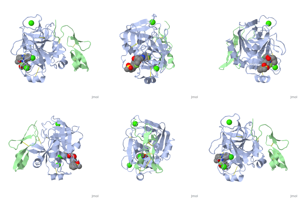
Factor X is cleaved by factor IXa (in the intrinsic pathway), or by factor VIIa (in the extrinsic pathway) to release the activation peptide, and yield the active factor Xa. The vitamin K-dependent, enzymatic carboxylation of some glutamate residues allows the modified protein to bind calcium via the GLA domain. Zymogen human factor X has four carbohydrate-attachment sites in the activation peptide, O-glycosidic linkages are to Thr17 and Thr29, and N-glycosidic linkages are to Asn39 and Asn49. | Factor X is cleaved by factor IXa (in the intrinsic pathway), or by factor VIIa (in the extrinsic pathway) to release the activation peptide, and yield the active factor Xa. The vitamin K-dependent, enzymatic carboxylation of some glutamate residues allows the modified protein to bind calcium via the GLA domain. Zymogen human factor X has four carbohydrate-attachment sites in the activation peptide, O-glycosidic linkages are to Thr17 and Thr29, and N-glycosidic linkages are to Asn39 and Asn49. | ||
</StructureSection> | </StructureSection> | ||
| - | __NOTOC__ | ||
==Enzyme Mechanism== | ==Enzyme Mechanism== | ||
| Line 69: | Line 68: | ||
<applet load='1GCI' size='300' frame='true' align='right' scene='Factor_Xa/Lbhb/1' caption='Possible LBHB between His57 and Asp102' /> | <applet load='1GCI' size='300' frame='true' align='right' scene='Factor_Xa/Lbhb/1' caption='Possible LBHB between His57 and Asp102' /> | ||
The mechanism by which the transition state is stabilized has been the topic of recent debate. Some groups suggest that His57 and Asp102 form and especially strong hydrogen bond, called a [http://en.wikipedia.org/wiki/Low-barrier_hydrogen_bond low barrier hydrogen bond (LBHB)]. They hypothesize that this hydrogen bond could promote formation of the transition state by stabilizing the Asp –His association and enhancing the bascisity of His57. <ref> PMID: 7661899</ref> <ref name="Frey alone">Frey, Perry A. Strong hydrogen bonding in chymotrypsin and other serine proteases. Journal of Physical Organic Chemistry (2004), 17(6-7), 511-520. </ref> This would enhance catalysis in the first step of the reaction. Formation of a LBHB requires a ΔpKa of approximately zero and a donor-to-acceptor distance of less then 2.65 Å for a nitrogen-oxygen pair like His57 and Asp102. Unlike a standard hydrogen bond, in which the hydrogen is located on the donor atom, a hydrogen in a LBHB is located equidistant between the 2 atoms. <ref name="subang"> PMID: 16834383 </ref> In 1998 Kuhn and colleagues published a crystal structure of ''Bacillus lentus'' subtilisn, another serine proetase, with 0.78 Å resolution at pH 5.9. The structure showed a distance of approximately 2.62 Å between the His57 nitrogen and the Asp102 oxygen, suggesting a LBHB. <ref> PMID: 9753430 </ref> | The mechanism by which the transition state is stabilized has been the topic of recent debate. Some groups suggest that His57 and Asp102 form and especially strong hydrogen bond, called a [http://en.wikipedia.org/wiki/Low-barrier_hydrogen_bond low barrier hydrogen bond (LBHB)]. They hypothesize that this hydrogen bond could promote formation of the transition state by stabilizing the Asp –His association and enhancing the bascisity of His57. <ref> PMID: 7661899</ref> <ref name="Frey alone">Frey, Perry A. Strong hydrogen bonding in chymotrypsin and other serine proteases. Journal of Physical Organic Chemistry (2004), 17(6-7), 511-520. </ref> This would enhance catalysis in the first step of the reaction. Formation of a LBHB requires a ΔpKa of approximately zero and a donor-to-acceptor distance of less then 2.65 Å for a nitrogen-oxygen pair like His57 and Asp102. Unlike a standard hydrogen bond, in which the hydrogen is located on the donor atom, a hydrogen in a LBHB is located equidistant between the 2 atoms. <ref name="subang"> PMID: 16834383 </ref> In 1998 Kuhn and colleagues published a crystal structure of ''Bacillus lentus'' subtilisn, another serine proetase, with 0.78 Å resolution at pH 5.9. The structure showed a distance of approximately 2.62 Å between the His57 nitrogen and the Asp102 oxygen, suggesting a LBHB. <ref> PMID: 9753430 </ref> | ||
| - | |||
| - | |||
A more recent crystal structure of α-Lytic protease, published in 2006 with 0.82 Å resolution argues against both the his flip mechanism and the presence of a LBHB between His57 and Asp102 (2.755 Å in this structure). Fuhrmann ''et al'' suggests that a LBHB may have been present in the subtilisin strucutre, it is not required for the serine protease mechanism. Instead they state that the chymotrypsin-like proteases may use a network of optimized hydrogen bonds to position the stabilize the tetrahedral intermediate and position the catalytic triad. Ser195 undergoes a shift of ~1Å upon protonation of His57 that destabilizes the His57-Ser195 H-bond. This conformation change would prevent His57 from reprotonating Ser195 leading to regeneration of the substrate.<ref name="subang" /> | A more recent crystal structure of α-Lytic protease, published in 2006 with 0.82 Å resolution argues against both the his flip mechanism and the presence of a LBHB between His57 and Asp102 (2.755 Å in this structure). Fuhrmann ''et al'' suggests that a LBHB may have been present in the subtilisin strucutre, it is not required for the serine protease mechanism. Instead they state that the chymotrypsin-like proteases may use a network of optimized hydrogen bonds to position the stabilize the tetrahedral intermediate and position the catalytic triad. Ser195 undergoes a shift of ~1Å upon protonation of His57 that destabilizes the His57-Ser195 H-bond. This conformation change would prevent His57 from reprotonating Ser195 leading to regeneration of the substrate.<ref name="subang" /> | ||
| Line 77: | Line 74: | ||
Updated on {{REVISIONDAY2}}-{{MONTHNAME|{{REVISIONMONTH}}}}-{{REVISIONYEAR}} | Updated on {{REVISIONDAY2}}-{{MONTHNAME|{{REVISIONMONTH}}}}-{{REVISIONYEAR}} | ||
| + | |||
| + | ===Factor X=== | ||
[[1whe]], [[1whf]] – bFX GLA + EGF-like domains – bovine – NMR<br /> | [[1whe]], [[1whf]] – bFX GLA + EGF-like domains – bovine – NMR<br /> | ||
Revision as of 07:07, 18 August 2014
| |||||||||||
Contents |
Enzyme Mechanism
General Serine Protease Mechanism
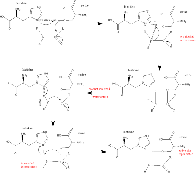
During the acylation half of the reaction His57 acts as a general base to remove a proton from Ser195, allowing it to attack the carbonyl of the peptide bond to be broken within the substrate, to yield the first tetrahedral intermediate. The negative oxygen ion of the tetrahedral intermediate is stabilized through hydrogen bonding with the oxyanion hole (Gly192 and Ser195). Asp102 stabilizes the protonated His57 through hydrogen bonding. His57 protonates the amine of the scissile bond, promoting formation of the acylenzyme and release of the N-terminal portion of the substrate.
The deacylation portion repeats the same sequence. A water molecule is deprotonated by His57 and attacks the acyl enzyme, to yielding a second tetrahedral intermediate. Again, the tetrahedral intermediate is stabilized by the oxyanion hole. Upon collapse of the tetrahedral intermediate, the C-terminal portion of the protein is released.[18]Controversial Mechanisms
His Flip Mechanisms

However, there are several arguments against the His flip mechanism. Flipping of His57 would require breaking and reforming many hydrogen bonds while the short lived tetrahedral intermediate is present. Also, His57 is sterically hindered by the P2 and P1’ residues of the peptide substrates. [23]These observations disfavor the His flip mechanism.
Low Barrier Hydrogen Bonds
|
The mechanism by which the transition state is stabilized has been the topic of recent debate. Some groups suggest that His57 and Asp102 form and especially strong hydrogen bond, called a low barrier hydrogen bond (LBHB). They hypothesize that this hydrogen bond could promote formation of the transition state by stabilizing the Asp –His association and enhancing the bascisity of His57. [24] [25] This would enhance catalysis in the first step of the reaction. Formation of a LBHB requires a ΔpKa of approximately zero and a donor-to-acceptor distance of less then 2.65 Å for a nitrogen-oxygen pair like His57 and Asp102. Unlike a standard hydrogen bond, in which the hydrogen is located on the donor atom, a hydrogen in a LBHB is located equidistant between the 2 atoms. [26] In 1998 Kuhn and colleagues published a crystal structure of Bacillus lentus subtilisn, another serine proetase, with 0.78 Å resolution at pH 5.9. The structure showed a distance of approximately 2.62 Å between the His57 nitrogen and the Asp102 oxygen, suggesting a LBHB. [27]
A more recent crystal structure of α-Lytic protease, published in 2006 with 0.82 Å resolution argues against both the his flip mechanism and the presence of a LBHB between His57 and Asp102 (2.755 Å in this structure). Fuhrmann et al suggests that a LBHB may have been present in the subtilisin strucutre, it is not required for the serine protease mechanism. Instead they state that the chymotrypsin-like proteases may use a network of optimized hydrogen bonds to position the stabilize the tetrahedral intermediate and position the catalytic triad. Ser195 undergoes a shift of ~1Å upon protonation of His57 that destabilizes the His57-Ser195 H-bond. This conformation change would prevent His57 from reprotonating Ser195 leading to regeneration of the substrate.[26]
3D structures of factor X
Updated on 18-August-2014
Factor X
1whe, 1whf – bFX GLA + EGF-like domains – bovine – NMR
1ccf, 1apo - bFX EGF-like domain – NMR
1hcg - hFX heavy chain catalytic domain + light chain EGF-like domain (residues 129-179) – human
1c5m - hFX heavy chain catalytic domain + light chain residues 84-179
Factor X complex with inhibitor
2ei6, 2ei7, 2ei8, 2p93, 2p94, 2p95, 2d1j, 1wu1, 1v3x – hFX heavy chain residues 16-243 + light chain EGF-like domain + inhibitor
2ra0, 2p3t, 2p16, 2p3u, 2pr3, 2q1j, 2g00, 2fzz, 2bq6, 2bq7, 2bqw, 1z6e, 2bmg, 2bok, 1mq5, 1mq6, 1fjs, 3k9x, 3ffg, 3kl6, 3hpt, 2vvc, 2vvu, 2vvv, 2vwl, 2vwm, 2vwn, 2vwo, 2w3i, 2w3k, 2jkh, 3ens, 2vh6, 2vh0, 2w26, 3cs7, 3cen, 2phb, 2y7x, 2y7z, 2y80, 2y81, 2y82, 2wyg, 2wyj, 2xbv, 2xbw, 2xbx, 2xby, 1ioe, 1iqe, 1iqf, 1iqg, 1iqh, 1iqi, 1iqj, 1iqk, 1iql, 1iqm, 1iqn, 1g2l, 1g2m, 1xka, 1xkb, 1fax, 2y5f, 2y5g, 2y5h, 3q3k, 4a7i, 3m36, 3m37, 3tk5, 3tk6 - hFX heavy chain catalytic domain + light chain EGF-like domain + inhibitor
2boh - hFX heavy chain catalytic domain (mutant) + light chain EGF-like domain (mutant) + inhibitor
2uwo, 2uwp, 2uwl, 2j94, 2j95, 2j2u, 2j34, 2j38, 2j4i, 2cji, 1lqd, 1lpg, 1lpk, 1lpz, 1ksn, 1ezq, 1f0r, 1f0s - hFX heavy chain catalytic domain + light chain GLA and EGF-like domains + inhibitor
1nfu, 1nfw, 1nfx, 1nfy - hFX heavy chain catalytic domain + light chain residues 46-240 + inhibitor
3sw2 - hFX heavy chain catalytic domain + light chain residues 84-179 + inhibitor
2xc0, 2xc4, 2xc5, 3iit, 3kqb, 3kqc, 3kqd, 3kqe, 3liw - hFX heavy chain catalytic domain + light chain EGF-like domain + inhibitor
4bti - hFX heavy chain + light chain + inhibitor
Factor X binary complex with protein
2h9e - hFX heavy chain catalytic domain + light chain residues 86-234 + anti-coagulant protein C2 + DTY-IRL-LPD
2p3f - hFX heavy chain catalytic domain + light chain EGF-like domain + anti-coagulant protein 5
2gd4 - hFX heavy chain catalytic domain (mutant) + light chain EGF-like domain + anti-thrombin III (mutant)
1p0s - hFX heavy chain catalytic domain + light chain GLA and EGF-like domains + ecotin precursor (mutant)
1iod – bFX GLA domain + FX-binding protein
1kig - bFX heavy chain catalytic domain + light chain EGF-like domain + anticoagulant peptide
1fxy – hFX/trypsin + PPR-CMK
4bxw – FX catalytic domain + coagulation factor V – brown snake
Additional Resources
For additional information, see: Hemophilia
References
- ↑ Cite error: Invalid
<ref>tag; no text was provided for refs namedGreer - ↑ 2.0 2.1 Department of Chemistry, University of Maine, Orono, ME. http://chemistry.umeche.maine.edu/CHY252/Peptidase3.html
- ↑ Padmanabhan K, Padmanabhan KP, Tulinsky A, Park CH, Bode W, Huber R, Blankenship DT, Cardin AD, Kisiel W. Structure of human des(1-45) factor Xa at 2.2 A resolution. J Mol Biol. 1993 Aug 5;232(3):947-66. PMID:8355279 doi:http://dx.doi.org/10.1006/jmbi.1993.1441
- ↑ Friedman PA, Przysiecki CT. Vitamin K-dependent carboxylation. Int J Biochem. 1987;19(1):1-7. PMID:3106112
- ↑ Vermeer C. Gamma-carboxyglutamate-containing proteins and the vitamin K-dependent carboxylase. Biochem J. 1990 Mar 15;266(3):625-36. PMID:2183788
- ↑ Price PA, Fraser JD, Metz-Virca G. Molecular cloning of matrix Gla protein: implications for substrate recognition by the vitamin K-dependent gamma-carboxylase. Proc Natl Acad Sci U S A. 1987 Dec;84(23):8335-9. PMID:3317405
- ↑ Freedman SJ, Furie BC, Furie B, Baleja JD. Structure of the metal-free gamma-carboxyglutamic acid-rich membrane binding region of factor IX by two-dimensional NMR spectroscopy. J Biol Chem. 1995 Apr 7;270(14):7980-7. PMID:7713897
- ↑ Freedman SJ, Blostein MD, Baleja JD, Jacobs M, Furie BC, Furie B. Identification of the phospholipid binding site in the vitamin K-dependent blood coagulation protein factor IX. J Biol Chem. 1996 Jul 5;271(27):16227-36. PMID:8663165
- ↑ Morita T, Jackson CM. Preparation and properties of derivatives of bovine factor X and factor Xa from which the gamma-carboxyglutamic acid containing domain has been removed. J Biol Chem. 1986 Mar 25;261(9):4015-23. PMID:3512564
- ↑ Muskavitch MA, Hoffmann FM. Homologs of vertebrate growth factors in Drosophila melanogaster and other invertebrates. Curr Top Dev Biol. 1990;24:289-328. PMID:2116263
- ↑ Ohlin AK, Linse S, Stenflo J. Calcium binding to the epidermal growth factor homology region of bovine protein C. J Biol Chem. 1988 May 25;263(15):7411-7. PMID:3259233
- ↑ Selander-Sunnerhagen M, Ullner M, Persson E, Teleman O, Stenflo J, Drakenberg T. How an epidermal growth factor (EGF)-like domain binds calcium. High resolution NMR structure of the calcium form of the NH2-terminal EGF-like domain in coagulation factor X. J Biol Chem. 1992 Sep 25;267(27):19642-9. PMID:1527084
- ↑ Persson E, Hogg PJ, Stenflo J. Effects of Ca2+ binding on the protease module of factor Xa and its interaction with factor Va. Evidence for two Gla-independent Ca(2+)-binding sites in factor Xa. J Biol Chem. 1993 Oct 25;268(30):22531-9. PMID:8226763
- ↑ Persson E, Selander M, Linse S, Drakenberg T, Ohlin AK, Stenflo J. Calcium binding to the isolated beta-hydroxyaspartic acid-containing epidermal growth factor-like domain of bovine factor X. J Biol Chem. 1989 Oct 5;264(28):16897-904. PMID:2789221
- ↑ Hopfner KP, Kopetzki E, Kresse GB, Bode W, Huber R, Engh RA. New enzyme lineages by subdomain shuffling. Proc Natl Acad Sci U S A. 1998 Aug 18;95(17):9813-8. PMID:9707558
- ↑ Factor X. Wikipedia
- ↑ Serine Protease. Wikipedia
- ↑ 18.0 18.1 Hedstrom L. Serine protease mechanism and specificity. Chem Rev. 2002 Dec;102(12):4501-24. PMID:12475199
- ↑ 19.0 19.1 Rai R, Sprengeler PA, Elrod KC, Young WB. Perspectives on factor Xa inhibition. Curr Med Chem. 2001 Feb;8(2):101-19. PMID:11172669
- ↑ www.bmolchem.wisc.edu/
- ↑ 21.0 21.1 Bachovchin, W. Contributions of NMR spectroscopy to the study of hydrogen bonds in serine protease active sites. Magnetic Resonance in Chemistry; (2001); 39(Spec. Issue); 199-213.
- ↑ Bachovchin WW. 15N NMR spectroscopy of hydrogen-bonding interactions in the active site of serine proteases: evidence for a moving histidine mechanism. Biochemistry. 1986 Nov 18;25(23):7751-9. PMID:3542033
- ↑ Brady K, Wei AZ, Ringe D, Abeles RH. Structure of chymotrypsin-trifluoromethyl ketone inhibitor complexes: comparison of slowly and rapidly equilibrating inhibitors. Biochemistry. 1990 Aug 21;29(33):7600-7. PMID:2271520
- ↑ Frey PA, Whitt SA, Tobin JB. A low-barrier hydrogen bond in the catalytic triad of serine proteases. Science. 1994 Jun 24;264(5167):1927-30. PMID:7661899
- ↑ Frey, Perry A. Strong hydrogen bonding in chymotrypsin and other serine proteases. Journal of Physical Organic Chemistry (2004), 17(6-7), 511-520.
- ↑ 26.0 26.1 Fuhrmann CN, Daugherty MD, Agard DA. Subangstrom crystallography reveals that short ionic hydrogen bonds, and not a His-Asp low-barrier hydrogen bond, stabilize the transition state in serine protease catalysis. J Am Chem Soc. 2006 Jul 19;128(28):9086-102. PMID:16834383 doi:http://dx.doi.org/10.1021/ja057721o
- ↑ Kuhn P, Knapp M, Soltis SM, Ganshaw G, Thoene M, Bott R. The 0.78 A structure of a serine protease: Bacillus lentus subtilisin. Biochemistry. 1998 Sep 29;37(39):13446-52. PMID:9753430 doi:10.1021/bi9813983
Proteopedia Page Contributors and Editors (what is this?)
Jacqueline Gertz, Michal Harel, Alexander Berchansky, David Canner, Jaime Prilusky


