This old version of Proteopedia is provided for student assignments while the new version is undergoing repairs. Content and edits done in this old version of Proteopedia after March 1, 2026 will eventually be lost when it is retired in about June of 2026.
Apply for new accounts at the new Proteopedia. Your logins will work in both the old and new versions.
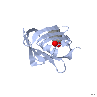
1cbs
From Proteopedia
(Difference between revisions)
| Line 1: | Line 1: | ||
| - | [[ | + | ==CRYSTAL STRUCTURE OF CELLULAR RETINOIC-ACID-BINDING PROTEINS I AND II IN COMPLEX WITH ALL-TRANS-RETINOIC ACID AND A SYNTHETIC RETINOID== |
| + | <StructureSection load='1cbs' size='340' side='right' caption='[[1cbs]], [[Resolution|resolution]] 1.80Å' scene=''> | ||
| + | == Structural highlights == | ||
| + | <table><tr><td colspan='2'>[[1cbs]] is a 1 chain structure with sequence from [http://en.wikipedia.org/wiki/Homo_sapiens Homo sapiens]. Full crystallographic information is available from [http://oca.weizmann.ac.il/oca-bin/ocashort?id=1CBS OCA]. For a <b>guided tour on the structure components</b> use [http://oca.weizmann.ac.il/oca-docs/fgij/fg.htm?mol=1CBS FirstGlance]. <br> | ||
| + | </td></tr><tr><td class="sblockLbl"><b>[[Ligand|Ligands:]]</b></td><td class="sblockDat"><scene name='pdbligand=REA:RETINOIC+ACID'>REA</scene><br> | ||
| + | <tr><td class="sblockLbl"><b>[[Gene|Gene:]]</b></td><td class="sblockDat">HUMAN CRABP-II ([http://www.ncbi.nlm.nih.gov/Taxonomy/Browser/wwwtax.cgi?mode=Info&srchmode=5&id=9606 Homo sapiens])</td></tr> | ||
| + | <tr><td class="sblockLbl"><b>Resources:</b></td><td class="sblockDat"><span class='plainlinks'>[http://oca.weizmann.ac.il/oca-docs/fgij/fg.htm?mol=1cbs FirstGlance], [http://oca.weizmann.ac.il/oca-bin/ocaids?id=1cbs OCA], [http://www.rcsb.org/pdb/explore.do?structureId=1cbs RCSB], [http://www.ebi.ac.uk/pdbsum/1cbs PDBsum]</span></td></tr> | ||
| + | <table> | ||
| + | == Evolutionary Conservation == | ||
| + | [[Image:Consurf_key_small.gif|200px|right]] | ||
| + | Check<jmol> | ||
| + | <jmolCheckbox> | ||
| + | <scriptWhenChecked>select protein; define ~consurf_to_do selected; consurf_initial_scene = true; script "/wiki/ConSurf/cb/1cbs_consurf.spt"</scriptWhenChecked> | ||
| + | <scriptWhenUnchecked>script /wiki/extensions/Proteopedia/spt/initialview01.spt</scriptWhenUnchecked> | ||
| + | <text>to colour the structure by Evolutionary Conservation</text> | ||
| + | </jmolCheckbox> | ||
| + | </jmol>, as determined by [http://consurfdb.tau.ac.il/ ConSurfDB]. You may read the [[Conservation%2C_Evolutionary|explanation]] of the method and the full data available from [http://bental.tau.ac.il/new_ConSurfDB/chain_selection.php?pdb_ID=2ata ConSurf]. | ||
| + | <div style="clear:both"></div> | ||
| + | <div style="background-color:#fffaf0;"> | ||
| + | == Publication Abstract from PubMed == | ||
| + | BACKGROUND: Retinoic acid (RA) plays a fundamental role in diverse cellular activities. Cellular RA binding proteins (CRABPs) are thought to act by modulating the amount of RA available to nuclear RA receptors. CRABPs and cellular retinol-binding proteins (CRBPs) share a unique fold of two orthogonal beta-sheets that encapsulate their ligands. It has been suggested that a trio of residues are the prime determinants defining the high specificity of CRBPs and CRABPs for their physiological ligands. RESULTS: Bovine/murine CRABP I and human CRABP II have been crystallized in complex with their natural ligand, all-trans-RA. Human CRABP II has also been crystallized in complex with a synthetic retinoid, 'compound 19'. Their structures have been determined and refined at resolutions of 2.9 A, 1.8 A and 2.2 A, respectively. CONCLUSIONS: The retinoid-binding site in CRABPs differs significantly from that observed in CRBP. Structural changes in three juxtaposed areas of the protein create a new, displaced binding site for RA. The carboxylate of the ligand interacts with the expected trio of residues (Arg132, Tyr134 and Arg111; CRABP II numbering). The RA ligand is almost flat with the beta-ionone ring showing a significant deviation (-33 degrees) from a cis conformation relative to the isoprene tail. The edge atoms of the beta-ionone ring are accessible to solvent in a suitable orientation for presentation to metabolizing enzymes. The bulkier synthetic retinoid causes small conformational changes in the protein structure. | ||
| - | + | Crystal structures of cellular retinoic acid binding proteins I and II in complex with all-trans-retinoic acid and a synthetic retinoid.,Kleywegt GJ, Bergfors T, Senn H, Le Motte P, Gsell B, Shudo K, Jones TA Structure. 1994 Dec 15;2(12):1241-58. PMID:7704533<ref>PMID:7704533</ref> | |
| - | + | From MEDLINE®/PubMed®, a database of the U.S. National Library of Medicine.<br> | |
| - | + | </div> | |
| - | + | ||
| - | + | ||
| - | + | ||
| - | + | ||
==See Also== | ==See Also== | ||
| Line 15: | Line 31: | ||
*[[Gustavo Elberto Epalza Sanchez/Sandbox 1|Gustavo Elberto Epalza Sanchez/Sandbox 1]] | *[[Gustavo Elberto Epalza Sanchez/Sandbox 1|Gustavo Elberto Epalza Sanchez/Sandbox 1]] | ||
*[[Molecular Playground/CRABP I (Cellular Retinoic Acid Binding Protein)|Molecular Playground/CRABP I (Cellular Retinoic Acid Binding Protein)]] | *[[Molecular Playground/CRABP I (Cellular Retinoic Acid Binding Protein)|Molecular Playground/CRABP I (Cellular Retinoic Acid Binding Protein)]] | ||
| - | + | == References == | |
| - | == | + | <references/> |
| - | < | + | __TOC__ |
| + | </StructureSection> | ||
[[Category: Homo sapiens]] | [[Category: Homo sapiens]] | ||
[[Category: Bergfors, T.]] | [[Category: Bergfors, T.]] | ||
Revision as of 17:05, 20 August 2014
CRYSTAL STRUCTURE OF CELLULAR RETINOIC-ACID-BINDING PROTEINS I AND II IN COMPLEX WITH ALL-TRANS-RETINOIC ACID AND A SYNTHETIC RETINOID
| |||||||||||