We apologize for Proteopedia being slow to respond. For the past two years, a new implementation of Proteopedia has been being built. Soon, it will replace this 18-year old system. All existing content will be moved to the new system at a date that will be announced here.
1lbj
From Proteopedia
(Difference between revisions)
| Line 1: | Line 1: | ||
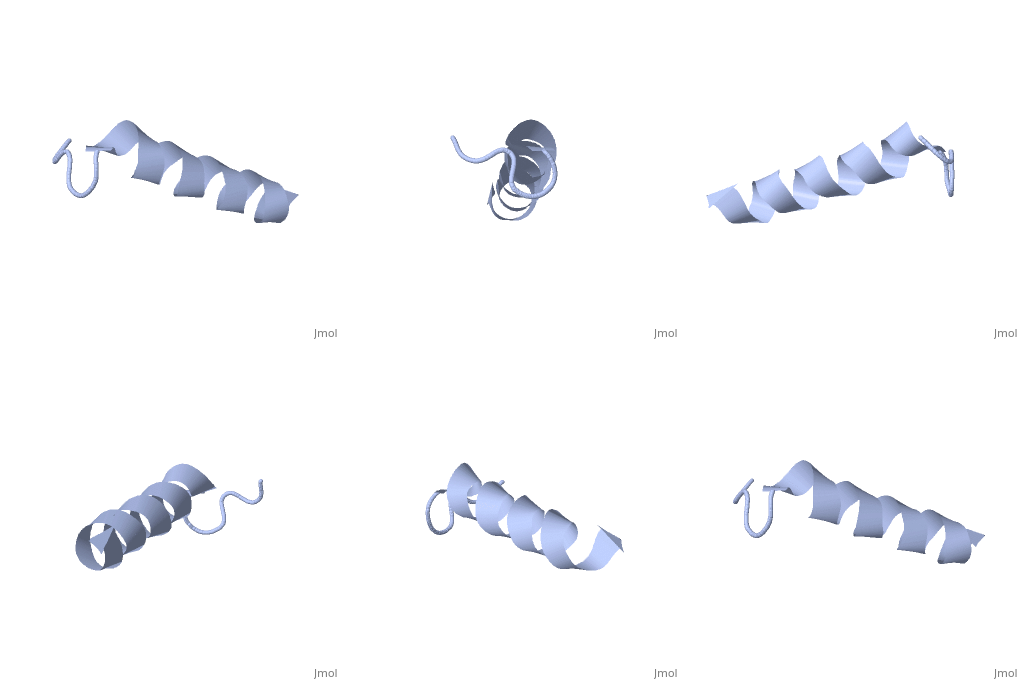
| - | [[ | + | ==NMR solution structure of motilin in phospholipid bicellar solution== |
| + | <StructureSection load='1lbj' size='340' side='right' caption='[[1lbj]], [[NMR_Ensembles_of_Models | 24 NMR models]]' scene=''> | ||
| + | == Structural highlights == | ||
| + | <table><tr><td colspan='2'>[[1lbj]] is a 1 chain structure. Full experimental information is available from [http://oca.weizmann.ac.il/oca-bin/ocashort?id=1LBJ OCA]. For a <b>guided tour on the structure components</b> use [http://oca.weizmann.ac.il/oca-docs/fgij/fg.htm?mol=1LBJ FirstGlance]. <br> | ||
| + | </td></tr><tr><td class="sblockLbl"><b>Resources:</b></td><td class="sblockDat"><span class='plainlinks'>[http://oca.weizmann.ac.il/oca-docs/fgij/fg.htm?mol=1lbj FirstGlance], [http://oca.weizmann.ac.il/oca-bin/ocaids?id=1lbj OCA], [http://www.rcsb.org/pdb/explore.do?structureId=1lbj RCSB], [http://www.ebi.ac.uk/pdbsum/1lbj PDBsum]</span></td></tr> | ||
| + | <table> | ||
| + | <div style="background-color:#fffaf0;"> | ||
| + | == Publication Abstract from PubMed == | ||
| + | The structure and dynamics of the gastrointestinal peptide hormone motilin, consisting of 22 amino acid residues, have been studied in the presence of isotropic q = 0.5 phospholipid bicelles. The NMR solution structure of the peptide in acidic bicelle solution was determined from 203 NOE-derived distance constraints and six backbone torsion angle constraints. Dynamic properties for the 13Calpha-1H vector in Leu10 were determined for motilin specifically labeled with 13C at this position by analysis of multiple-field relaxation data. The structure reveals an ordered alpha-helical conformation between Glu9 and Lys20. The N-terminus is also well structured with a turn resembling that of a classical beta-turn. The 13C dynamics clearly show that motilin tumbles slowly in solution, with a correlation time characteristic of a large object. It was also found that motilin has a large degree of local flexibility as compared with what has previously been reported in SDS micelles. The results show that motilin interacts with the bicelle, displaying motional properties of a peptide bound to a membrane. In comparison, motilin in neutral bicelles seems less structured and more flexible. This study shows that the small isotropic bicelles are well suited for use as membrane-mimetic for structural as well as dynamical investigations of membrane-bound peptides by high-resolution NMR. | ||
| - | + | NMR solution structure and dynamics of motilin in isotropic phospholipid bicellar solution.,Andersson A, Maler L J Biomol NMR. 2002 Oct;24(2):103-12. PMID:12495026<ref>PMID:12495026</ref> | |
| - | + | From MEDLINE®/PubMed®, a database of the U.S. National Library of Medicine.<br> | |
| - | + | </div> | |
| - | + | ||
| - | + | ||
| - | + | ||
| - | + | ||
==See Also== | ==See Also== | ||
*[[Motilin|Motilin]] | *[[Motilin|Motilin]] | ||
| - | + | == References == | |
| - | == | + | <references/> |
| - | < | + | __TOC__ |
| + | </StructureSection> | ||
[[Category: Andersson, A.]] | [[Category: Andersson, A.]] | ||
[[Category: Maler, L.]] | [[Category: Maler, L.]] | ||
Revision as of 16:54, 28 September 2014
NMR solution structure of motilin in phospholipid bicellar solution
| |||||||||||

