This old version of Proteopedia is provided for student assignments while the new version is undergoing repairs. Content and edits done in this old version of Proteopedia after March 1, 2026 will eventually be lost when it is retired in about June of 2026.
Apply for new accounts at the new Proteopedia. Your logins will work in both the old and new versions.
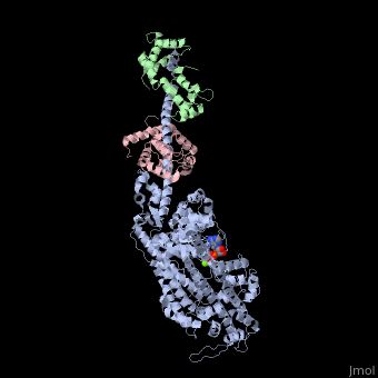
3i5f
From Proteopedia
| Line 1: | Line 1: | ||
| - | [[ | + | ==Crystal structure of squid MG.ADP myosin S1== |
| + | <StructureSection load='3i5f' size='340' side='right' caption='[[3i5f]], [[Resolution|resolution]] 3.10Å' scene=''> | ||
| + | == Structural highlights == | ||
| + | <table><tr><td colspan='2'>[[3i5f]] is a 3 chain structure with sequence from [http://en.wikipedia.org/wiki/Doryteuthis_pealeii Doryteuthis pealeii] and [http://en.wikipedia.org/wiki/Todarodes_pacificus Todarodes pacificus]. This structure supersedes the now removed PDB entry [http://oca.weizmann.ac.il/oca-bin/send-pdb?obs=1&id=2oy6 2oy6]. Full crystallographic information is available from [http://oca.weizmann.ac.il/oca-bin/ocashort?id=3I5F OCA]. For a <b>guided tour on the structure components</b> use [http://oca.weizmann.ac.il/oca-docs/fgij/fg.htm?mol=3I5F FirstGlance]. <br> | ||
| + | </td></tr><tr><td class="sblockLbl"><b>[[Ligand|Ligands:]]</b></td><td class="sblockDat"><scene name='pdbligand=ADP:ADENOSINE-5-DIPHOSPHATE'>ADP</scene>, <scene name='pdbligand=MG:MAGNESIUM+ION'>MG</scene><br> | ||
| + | <tr><td class="sblockLbl"><b>[[Related_structure|Related:]]</b></td><td class="sblockDat">[[2oy6|2oy6]], [[3i5g|3i5g]], [[3i5h|3i5h]], [[3i5i|3i5i]]</td></tr> | ||
| + | <tr><td class="sblockLbl"><b>Resources:</b></td><td class="sblockDat"><span class='plainlinks'>[http://oca.weizmann.ac.il/oca-docs/fgij/fg.htm?mol=3i5f FirstGlance], [http://oca.weizmann.ac.il/oca-bin/ocaids?id=3i5f OCA], [http://www.rcsb.org/pdb/explore.do?structureId=3i5f RCSB], [http://www.ebi.ac.uk/pdbsum/3i5f PDBsum]</span></td></tr> | ||
| + | <table> | ||
| + | == Evolutionary Conservation == | ||
| + | [[Image:Consurf_key_small.gif|200px|right]] | ||
| + | Check<jmol> | ||
| + | <jmolCheckbox> | ||
| + | <scriptWhenChecked>select protein; define ~consurf_to_do selected; consurf_initial_scene = true; script "/wiki/ConSurf/i5/3i5f_consurf.spt"</scriptWhenChecked> | ||
| + | <scriptWhenUnchecked>script /wiki/extensions/Proteopedia/spt/initialview01.spt</scriptWhenUnchecked> | ||
| + | <text>to colour the structure by Evolutionary Conservation</text> | ||
| + | </jmolCheckbox> | ||
| + | </jmol>, as determined by [http://consurfdb.tau.ac.il/ ConSurfDB]. You may read the [[Conservation%2C_Evolutionary|explanation]] of the method and the full data available from [http://bental.tau.ac.il/new_ConSurfDB/chain_selection.php?pdb_ID=2ata ConSurf]. | ||
| + | <div style="clear:both"></div> | ||
| + | <div style="background-color:#fffaf0;"> | ||
| + | == Publication Abstract from PubMed == | ||
| + | Unlike processive cellular motors such as myosin V, whose structure has recently been determined in a "rigor-like" conformation, myosin II from contracting muscle filaments necessarily spends most of its time detached from actin. By using squid and sea scallop sources, however, we have now obtained similar rigor-like atomic structures for muscle myosin heads (S1). The significance of the hallmark closed actin-binding cleft in these crystal structures is supported here by actin/S1-binding studies. These structures reveal how different duty ratios, and hence cellular functions, of the myosin isoforms may be accounted for, in part, on the basis of detailed differences in interdomain contacts. Moreover, the rigor-like position of switch II turns out to be unique for myosin V. The overall arrangements of subdomains in the motor are relatively conserved in each of the known contractile states, and we explore qualitatively the energetics of these states. | ||
| - | + | Rigor-like structures from muscle myosins reveal key mechanical elements in the transduction pathways of this allosteric motor.,Yang Y, Gourinath S, Kovacs M, Nyitray L, Reutzel R, Himmel DM, O'Neall-Hennessey E, Reshetnikova L, Szent-Gyorgyi AG, Brown JH, Cohen C Structure. 2007 May;15(5):553-64. PMID:17502101<ref>PMID:17502101</ref> | |
| - | + | From MEDLINE®/PubMed®, a database of the U.S. National Library of Medicine.<br> | |
| - | + | </div> | |
| - | + | ||
| - | + | ||
| - | + | ||
| - | + | ||
==See Also== | ==See Also== | ||
*[[Myosin|Myosin]] | *[[Myosin|Myosin]] | ||
| - | + | == References == | |
| - | == | + | <references/> |
| - | < | + | __TOC__ |
| + | </StructureSection> | ||
[[Category: Doryteuthis pealeii]] | [[Category: Doryteuthis pealeii]] | ||
[[Category: Todarodes pacificus]] | [[Category: Todarodes pacificus]] | ||
Revision as of 13:08, 29 September 2014
Crystal structure of squid MG.ADP myosin S1
| |||||||||||

Categories: Doryteuthis pealeii | Todarodes pacificus | Brown, J H. | Cohen, C. | Gourinath, S. | Himmel, D M. | Kovacs, M. | Neall-Hennessey, E O. | Nyitray, L. | Reshetnikova, L. | Reutzel, R. | Szent-Gyorgyi, A G. | Yang, Y. | Actin-binding | Atp-binding | Contractile protein | Mg adp | Motor protein | Muscle protein | Myosin | Myosin ii s1 | Nucleotide-binding | Post-rigor state | Squid | Thick filament
