This old version of Proteopedia is provided for student assignments while the new version is undergoing repairs. Content and edits done in this old version of Proteopedia after March 1, 2026 will eventually be lost when it is retired in about June of 2026.
Apply for new accounts at the new Proteopedia. Your logins will work in both the old and new versions.
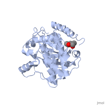
1j3h
From Proteopedia
(Difference between revisions)
| Line 1: | Line 1: | ||
| - | + | ==Crystal structure of apoenzyme cAMP-dependent protein kinase catalytic subunit== | |
| - | === | + | <StructureSection load='1j3h' size='340' side='right' caption='[[1j3h]], [[Resolution|resolution]] 2.90Å' scene=''> |
| - | + | == Structural highlights == | |
| + | <table><tr><td colspan='2'>[[1j3h]] is a 2 chain structure with sequence from [http://en.wikipedia.org/wiki/Mus_musculus Mus musculus]. Full crystallographic information is available from [http://oca.weizmann.ac.il/oca-bin/ocashort?id=1J3H OCA]. For a <b>guided tour on the structure components</b> use [http://oca.weizmann.ac.il/oca-docs/fgij/fg.htm?mol=1J3H FirstGlance]. <br> | ||
| + | </td></tr><tr><td class="sblockLbl"><b>[[Ligand|Ligands:]]</b></td><td class="sblockDat"><scene name='pdbligand=MPD:(4S)-2-METHYL-2,4-PENTANEDIOL'>MPD</scene><br> | ||
| + | <tr><td class="sblockLbl"><b>[[Non-Standard_Residue|NonStd Res:]]</b></td><td class="sblockDat"><scene name='pdbligand=CME:S,S-(2-HYDROXYETHYL)THIOCYSTEINE'>CME</scene>, <scene name='pdbligand=SEP:PHOSPHOSERINE'>SEP</scene>, <scene name='pdbligand=TPO:PHOSPHOTHREONINE'>TPO</scene></td></tr> | ||
| + | <tr><td class="sblockLbl"><b>[[Related_structure|Related:]]</b></td><td class="sblockDat">[[1l3r|1l3r]], [[1ctp|1ctp]], [[1cmk|1cmk]], [[1atp|1atp]], [[1bkx|1bkx]]</td></tr> | ||
| + | <tr><td class="sblockLbl"><b>Activity:</b></td><td class="sblockDat"><span class='plainlinks'>[http://en.wikipedia.org/wiki/Non-specific_serine/threonine_protein_kinase Non-specific serine/threonine protein kinase], with EC number [http://www.brenda-enzymes.info/php/result_flat.php4?ecno=2.7.11.1 2.7.11.1] </span></td></tr> | ||
| + | <tr><td class="sblockLbl"><b>Resources:</b></td><td class="sblockDat"><span class='plainlinks'>[http://oca.weizmann.ac.il/oca-docs/fgij/fg.htm?mol=1j3h FirstGlance], [http://oca.weizmann.ac.il/oca-bin/ocaids?id=1j3h OCA], [http://www.rcsb.org/pdb/explore.do?structureId=1j3h RCSB], [http://www.ebi.ac.uk/pdbsum/1j3h PDBsum]</span></td></tr> | ||
| + | <table> | ||
| + | == Evolutionary Conservation == | ||
| + | [[Image:Consurf_key_small.gif|200px|right]] | ||
| + | Check<jmol> | ||
| + | <jmolCheckbox> | ||
| + | <scriptWhenChecked>select protein; define ~consurf_to_do selected; consurf_initial_scene = true; script "/wiki/ConSurf/j3/1j3h_consurf.spt"</scriptWhenChecked> | ||
| + | <scriptWhenUnchecked>script /wiki/extensions/Proteopedia/spt/initialview01.spt</scriptWhenUnchecked> | ||
| + | <text>to colour the structure by Evolutionary Conservation</text> | ||
| + | </jmolCheckbox> | ||
| + | </jmol>, as determined by [http://consurfdb.tau.ac.il/ ConSurfDB]. You may read the [[Conservation%2C_Evolutionary|explanation]] of the method and the full data available from [http://bental.tau.ac.il/new_ConSurfDB/chain_selection.php?pdb_ID=2ata ConSurf]. | ||
| + | <div style="clear:both"></div> | ||
| + | <div style="background-color:#fffaf0;"> | ||
| + | == Publication Abstract from PubMed == | ||
| + | To better understand the mechanism of ligand binding and ligand-induced conformational change, the crystal structure of apoenzyme catalytic (C) subunit of adenosine-3',5'-cyclic monophosphate (cAMP)-dependent protein kinase (PKA) was solved. The apoenzyme structure (Apo) provides a snapshot of the enzyme in the first step of the catalytic cycle, and in this unliganded form the PKA C subunit adopts an open conformation. A hydrophobic junction is formed by residues from the small and large lobes that come into close contact. This "greasy" patch may lubricate the shearing motion associated with domain rotation, and the opening and closing of the active-site cleft. Although Apo appears to be quite dynamic, many important residues for MgATP binding and phosphoryl transfer in the active site are preformed. Residues around the adenine ring of ATP and residues involved in phosphoryl transfer from the large lobe are mostly preformed, whereas residues involved in ribose binding and in the Gly-rich loop are not. Prior to ligand binding, Lys72 and the C-terminal tail, two important ATP-binding elements are also disordered. The surface created in the active site is contoured to bind ATP, but not GTP, and appears to be held in place by a stable hydrophobic core, which includes helices C, E, and F, and beta strand 6. This core seems to provide a network for communicating from the active site, where nucleotide binds, to the peripheral peptide-binding F-to-G helix loop, exemplified by Phe239. Two potential lines of communication are the D helix and the F helix. The conserved Trp222-Phe238 network, which lies adjacent to the F-to-G helix loop, suggests that this network would exist in other protein kinases and may be a conserved means of communicating ATP binding from the active site to the distal peptide-binding ledge. | ||
| - | + | Dynamic features of cAMP-dependent protein kinase revealed by apoenzyme crystal structure.,Akamine P, Madhusudan, Wu J, Xuong NH, Ten Eyck LF, Taylor SS J Mol Biol. 2003 Mar 14;327(1):159-71. PMID:12614615<ref>PMID:12614615</ref> | |
| - | + | ||
| + | From MEDLINE®/PubMed®, a database of the U.S. National Library of Medicine.<br> | ||
| + | </div> | ||
==See Also== | ==See Also== | ||
*[[CAMP Dependent Protein Kinase%2C Catalytic Subunit|CAMP Dependent Protein Kinase%2C Catalytic Subunit]] | *[[CAMP Dependent Protein Kinase%2C Catalytic Subunit|CAMP Dependent Protein Kinase%2C Catalytic Subunit]] | ||
*[[CAMP-dependent protein kinase|CAMP-dependent protein kinase]] | *[[CAMP-dependent protein kinase|CAMP-dependent protein kinase]] | ||
| - | + | *[[Eukaryotic Protein Kinase Catalytic Domain|Eukaryotic Protein Kinase Catalytic Domain]] | |
| - | == | + | == References == |
| - | < | + | <references/> |
| + | __TOC__ | ||
| + | </StructureSection> | ||
[[Category: Mus musculus]] | [[Category: Mus musculus]] | ||
[[Category: Non-specific serine/threonine protein kinase]] | [[Category: Non-specific serine/threonine protein kinase]] | ||
Revision as of 16:03, 29 September 2014
Crystal structure of apoenzyme cAMP-dependent protein kinase catalytic subunit
| |||||||||||