We apologize for Proteopedia being slow to respond. For the past two years, a new implementation of Proteopedia has been being built. Soon, it will replace this 18-year old system. All existing content will be moved to the new system at a date that will be announced here.
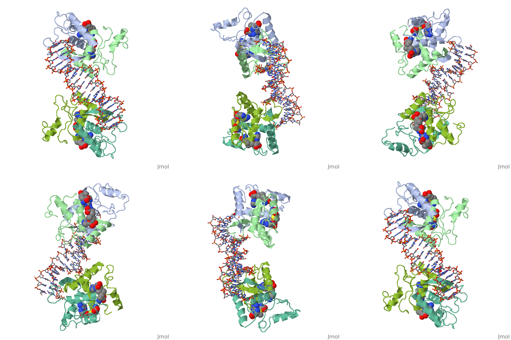
1cma
From Proteopedia
(Difference between revisions)
| Line 1: | Line 1: | ||
| - | + | ==MET REPRESSOR/DNA COMPLEX + S-ADENOSYL-METHIONINE== | |
| - | + | <StructureSection load='1cma' size='340' side='right' caption='[[1cma]], [[Resolution|resolution]] 2.80Å' scene=''> | |
| - | + | == Structural highlights == | |
| + | <table><tr><td colspan='2'>[[1cma]] is a 4 chain structure with sequence from [http://en.wikipedia.org/wiki/Escherichia_coli Escherichia coli]. Full crystallographic information is available from [http://oca.weizmann.ac.il/oca-bin/ocashort?id=1CMA OCA]. For a <b>guided tour on the structure components</b> use [http://oca.weizmann.ac.il/oca-docs/fgij/fg.htm?mol=1CMA FirstGlance]. <br> | ||
| + | </td></tr><tr><td class="sblockLbl"><b>[[Ligand|Ligands:]]</b></td><td class="sblockDat"><scene name='pdbligand=SAM:S-ADENOSYLMETHIONINE'>SAM</scene><br> | ||
| + | <tr><td class="sblockLbl"><b>Resources:</b></td><td class="sblockDat"><span class='plainlinks'>[http://oca.weizmann.ac.il/oca-docs/fgij/fg.htm?mol=1cma FirstGlance], [http://oca.weizmann.ac.il/oca-bin/ocaids?id=1cma OCA], [http://www.rcsb.org/pdb/explore.do?structureId=1cma RCSB], [http://www.ebi.ac.uk/pdbsum/1cma PDBsum]</span></td></tr> | ||
| + | <table> | ||
| + | == Evolutionary Conservation == | ||
| + | [[Image:Consurf_key_small.gif|200px|right]] | ||
| + | Check<jmol> | ||
| + | <jmolCheckbox> | ||
| + | <scriptWhenChecked>select protein; define ~consurf_to_do selected; consurf_initial_scene = true; script "/wiki/ConSurf/cm/1cma_consurf.spt"</scriptWhenChecked> | ||
| + | <scriptWhenUnchecked>script /wiki/extensions/Proteopedia/spt/initialview01.spt</scriptWhenUnchecked> | ||
| + | <text>to colour the structure by Evolutionary Conservation</text> | ||
| + | </jmolCheckbox> | ||
| + | </jmol>, as determined by [http://consurfdb.tau.ac.il/ ConSurfDB]. You may read the [[Conservation%2C_Evolutionary|explanation]] of the method and the full data available from [http://bental.tau.ac.il/new_ConSurfDB/chain_selection.php?pdb_ID=2ata ConSurf]. | ||
| + | <div style="clear:both"></div> | ||
| + | <div style="background-color:#fffaf0;"> | ||
| + | == Publication Abstract from PubMed == | ||
| + | The crystal structure of the met repressor-operator complex shows two dimeric repressor molecules bound to adjacent sites 8 base pairs apart on an 18-base-pair DNA fragment. Sequence specificity is achieved by insertion of double-stranded antiparallel protein beta-ribbons into the major groove of B-form DNA, with direct hydrogen-bonding between amino-acid side chains and the base pairs. The repressor also recognizes sequence-dependent distortion or flexibility of the operator phosphate backbone, conferring specificity even for inaccessible base pairs. | ||
| - | + | Crystal structure of the met repressor-operator complex at 2.8 A resolution reveals DNA recognition by beta-strands.,Somers WS, Phillips SE Nature. 1992 Oct 1;359(6394):387-93. PMID:1406951<ref>PMID:1406951</ref> | |
| - | + | ||
| + | From MEDLINE®/PubMed®, a database of the U.S. National Library of Medicine.<br> | ||
| + | </div> | ||
==See Also== | ==See Also== | ||
*[[Joe Granger Methionine Repressor: Escherichia coli|Joe Granger Methionine Repressor: Escherichia coli]] | *[[Joe Granger Methionine Repressor: Escherichia coli|Joe Granger Methionine Repressor: Escherichia coli]] | ||
| - | + | *[[Met repressor|Met repressor]] | |
| - | == | + | == References == |
| - | < | + | <references/> |
| + | __TOC__ | ||
| + | </StructureSection> | ||
[[Category: Escherichia coli]] | [[Category: Escherichia coli]] | ||
[[Category: Phillips, S E.V.]] | [[Category: Phillips, S E.V.]] | ||
Revision as of 17:12, 29 September 2014
MET REPRESSOR/DNA COMPLEX + S-ADENOSYL-METHIONINE
| |||||||||||


