We apologize for Proteopedia being slow to respond. For the past two years, a new implementation of Proteopedia has been being built. Soon, it will replace this 18-year old system. All existing content will be moved to the new system at a date that will be announced here.
Pheromone binding protein
From Proteopedia
(Difference between revisions)
| Line 18: | Line 18: | ||
== Structure == | == Structure == | ||
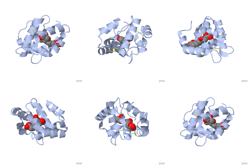
| - | The protein has a | + | The protein has a 1 alpha chain, that can be seen in pink. |
== Function == | == Function == | ||
| - | + | The protein ligand, 9-ODA [[Image:Example.jpg]] | |
== Structural highlights == | == Structural highlights == | ||
Revision as of 14:29, 19 November 2014
Introduction
| |||||||||||
References
- ↑ Pesenti ME, Spinelli S, Bezirard V, Briand L, Pernollet JC, Tegoni M, Cambillau C. Structural basis of the honey bee PBP pheromone and pH-induced conformational change. J Mol Biol. 2008 Jun 27;380(1):158-69. Epub 2008 Apr 27. PMID:18508083 doi:10.1016/j.jmb.2008.04.048
- ↑ Herraez A. Biomolecules in the computer: Jmol to the rescue. Biochem Mol Biol Educ. 2006 Jul;34(4):255-61. doi: 10.1002/bmb.2006.494034042644. PMID:21638687 doi:10.1002/bmb.2006.494034042644


