This old version of Proteopedia is provided for student assignments while the new version is undergoing repairs. Content and edits done in this old version of Proteopedia after March 1, 2026 will eventually be lost when it is retired in about June of 2026.
Apply for new accounts at the new Proteopedia. Your logins will work in both the old and new versions.
2zwh
From Proteopedia
(Difference between revisions)
| Line 1: | Line 1: | ||
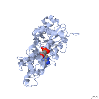
| - | + | ==Model for the F-actin structure== | |
| - | === | + | <StructureSection load='2zwh' size='340' side='right' caption='[[2zwh]], [[Resolution|resolution]] 3.30Å' scene=''> |
| - | + | == Structural highlights == | |
| + | <table><tr><td colspan='2'>[[2zwh]] is a 1 chain structure with sequence from [http://en.wikipedia.org/wiki/Oryctolagus_cuniculus Oryctolagus cuniculus]. Full crystallographic information is available from [http://oca.weizmann.ac.il/oca-bin/ocashort?id=2ZWH OCA]. For a <b>guided tour on the structure components</b> use [http://oca.weizmann.ac.il/oca-docs/fgij/fg.htm?mol=2ZWH FirstGlance]. <br> | ||
| + | </td></tr><tr id='ligand'><td class="sblockLbl"><b>[[Ligand|Ligands:]]</b></td><td class="sblockDat"><scene name='pdbligand=ADP:ADENOSINE-5-DIPHOSPHATE'>ADP</scene>, <scene name='pdbligand=CA:CALCIUM+ION'>CA</scene></td></tr> | ||
| + | <tr id='NonStdRes'><td class="sblockLbl"><b>[[Non-Standard_Residue|NonStd Res:]]</b></td><td class="sblockDat"><scene name='pdbligand=HIC:4-METHYL-HISTIDINE'>HIC</scene></td></tr> | ||
| + | <tr id='resources'><td class="sblockLbl"><b>Resources:</b></td><td class="sblockDat"><span class='plainlinks'>[http://oca.weizmann.ac.il/oca-docs/fgij/fg.htm?mol=2zwh FirstGlance], [http://oca.weizmann.ac.il/oca-bin/ocaids?id=2zwh OCA], [http://www.rcsb.org/pdb/explore.do?structureId=2zwh RCSB], [http://www.ebi.ac.uk/pdbsum/2zwh PDBsum]</span></td></tr> | ||
| + | </table> | ||
| + | <div style="background-color:#fffaf0;"> | ||
| + | == Publication Abstract from PubMed == | ||
| + | Actin plays crucial parts in cell motility through a dynamic process driven by polymerization and depolymerization, that is, the globular (G) to fibrous (F) actin transition. Although our knowledge about the actin-based cellular functions and the molecules that regulate the G- to F-actin transition is growing, the structural aspects of the transition remain enigmatic. We created a model of F-actin using X-ray fibre diffraction intensities obtained from well oriented sols of rabbit skeletal muscle F-actin to 3.3 A in the radial direction and 5.6 A along the equator. Here we show that the G- to F-actin conformational transition is a simple relative rotation of the two major domains by about 20 degrees. As a result of the domain rotation, the actin molecule in the filament is flat. The flat form is essential for the formation of stable, helical F-actin. Our F-actin structure model provides the basis for understanding actin polymerization as well as its molecular interactions with actin-binding proteins. | ||
| - | + | The nature of the globular- to fibrous-actin transition.,Oda T, Iwasa M, Aihara T, Maeda Y, Narita A Nature. 2009 Jan 22;457(7228):441-5. PMID:19158791<ref>PMID:19158791</ref> | |
| - | + | ||
| + | From MEDLINE®/PubMed®, a database of the U.S. National Library of Medicine.<br> | ||
| + | </div> | ||
==See Also== | ==See Also== | ||
*[[Actin|Actin]] | *[[Actin|Actin]] | ||
*[[F-actin|F-actin]] | *[[F-actin|F-actin]] | ||
| - | + | == References == | |
| - | == | + | <references/> |
| - | < | + | __TOC__ |
| + | </StructureSection> | ||
[[Category: Oryctolagus cuniculus]] | [[Category: Oryctolagus cuniculus]] | ||
| - | [[Category: Aihara, T | + | [[Category: Aihara, T]] |
| - | [[Category: Iwasa, M | + | [[Category: Iwasa, M]] |
| - | [[Category: Maeda, Y | + | [[Category: Maeda, Y]] |
| - | [[Category: Narita, A | + | [[Category: Narita, A]] |
| - | [[Category: Oda, T | + | [[Category: Oda, T]] |
[[Category: Atp-binding]] | [[Category: Atp-binding]] | ||
[[Category: Contractile protein]] | [[Category: Contractile protein]] | ||
Revision as of 14:02, 17 December 2014
Model for the F-actin structure
| |||||||||||