Sandbox CYPMetabolism
From Proteopedia
| Line 15: | Line 15: | ||
== The Heme Group is Vital for the Enzyme's Action == | == The Heme Group is Vital for the Enzyme's Action == | ||
| - | Let's look at the heme now. Click the following link to hide the rest of the protein and focus on the<scene name='60/609993/Cyp_1a2/2'>heme</scene> by itself. Carbon atoms are shown in grey, nitrogen in blue, oxygen in red, and the central iron atom in orange. The Iron atom is a vital center for oxidation of substrates (drugs or other xenobiotics). Rotate and re-size as necessary to shown that the heme is highly planar. Do you recognize the 4 pyrrole rings within the heme? | + | Let's look at the heme now. Click the following link to hide the rest of the protein and focus on the <scene name='60/609993/Cyp_1a2/2'>heme</scene> by itself. Carbon atoms are shown in grey, nitrogen in blue, oxygen in red, and the central iron atom in orange. The Iron atom is a vital center for oxidation of substrates (drugs or other xenobiotics). Rotate and re-size as necessary to shown that the heme is highly planar. Do you recognize the 4 pyrrole rings within the heme? |
Examine the iron atom in the heme. It is capable of forming 6 bonds. Four of those are with the 4 pyrrole rings that make up the porphyrin ring system. The fifth and sixth bonds are made to atoms above and below the plane of heme ring. The fifth is made to a cysteine residue present on the protein. The final bond is made to an oxygen molecule (not shown). This molecular oxygen is activated to aid in oxidation of substrates and would appear directly between the substrate and the iron atom. | Examine the iron atom in the heme. It is capable of forming 6 bonds. Four of those are with the 4 pyrrole rings that make up the porphyrin ring system. The fifth and sixth bonds are made to atoms above and below the plane of heme ring. The fifth is made to a cysteine residue present on the protein. The final bond is made to an oxygen molecule (not shown). This molecular oxygen is activated to aid in oxidation of substrates and would appear directly between the substrate and the iron atom. | ||
| Line 42: | Line 42: | ||
== Amino Acids in the Binding Pocket Affect Drug Selectivity == | == Amino Acids in the Binding Pocket Affect Drug Selectivity == | ||
| - | First examine the shape of the <scene name='60/609993/Cyp_1a2/9'>Van der Waals area</scene>around the flavone. The range of bluish-green to reddish-orange colored areas on the surface differentiate how close(red), or how far (blue), in proximity the flavone is to the binding pocket. These contacts can be caused by ionic, hydrophobic, or hydrogen bonding. Given the structure of the flavone, what forces do you suspect may be responsible for binding the enzyme so tightly? | + | First examine the shape of the <scene name='60/609993/Cyp_1a2/9'>Van der Waals area</scene> around the flavone. The range of bluish-green to reddish-orange colored areas on the surface differentiate how close(red), or how far (blue), in proximity the flavone is to the binding pocket. These contacts can be caused by ionic, hydrophobic, or hydrogen bonding. Given the structure of the flavone, what forces do you suspect may be responsible for binding the enzyme so tightly? |
In the next scene, the Van der Waals surface of the <scene name='60/609993/Cyp_1a2/21'>cavity</scene> is displayed. The portions of the cavity involved in binding are shown as orange patches. These are a result of specific amino acid residues that form the surface of the binding pocket. Clicking on this <scene name='60/609993/Cyp_1a2/20'>link</scene> will show the surface of the flavone and a few of the most important amino acid residues responsible for binding. | In the next scene, the Van der Waals surface of the <scene name='60/609993/Cyp_1a2/21'>cavity</scene> is displayed. The portions of the cavity involved in binding are shown as orange patches. These are a result of specific amino acid residues that form the surface of the binding pocket. Clicking on this <scene name='60/609993/Cyp_1a2/20'>link</scene> will show the surface of the flavone and a few of the most important amino acid residues responsible for binding. | ||
| Line 70: | Line 70: | ||
There are multiple types of interactions when considering CYP systems. Competitive inhibition follows fairly predictable rules. In this case, two drugs are competing for the same active site and one drug slows down the metabolism of the other. However, some drugs metabolized by CYP3A4 are known to have more complex interactions. | There are multiple types of interactions when considering CYP systems. Competitive inhibition follows fairly predictable rules. In this case, two drugs are competing for the same active site and one drug slows down the metabolism of the other. However, some drugs metabolized by CYP3A4 are known to have more complex interactions. | ||
| - | It has been demonstrated that two or more smaller molecules may bind within an active site at the same time. In this case, drug metabolism can, strangely enough, actually be increased. It has been proposed that when only one molecule of a smaller drug (let's call this Drug A) is bound to the active site, that the extent of Drug A 's metabolism can be minimal due to the relatively large cavity. One explanation may be that Drug A isn't held in a sufficient orientation to the heme Iron. For optimum metabolism, the heme should bond with an Oxygen of the ligand. However, when two molecules of a smaller drug bind at the same time, one molecule may help in forcing the other molecule to retain proper orientation; thus, improving catalytic efficiency. This theory of two drugs binding simultaneously to a CYP may influence its pharmacokinetics is illustrated by the following structure of ketoconazole bound to a CYP enzyme ([[2v0m]]) | + | It has been demonstrated that two or more smaller molecules may bind within an active site at the same time. In this case, drug metabolism can, strangely enough, actually be increased. It has been proposed that when only one molecule of a smaller drug (let's call this Drug A) is bound to the active site, that the extent of Drug A 's metabolism can be minimal due to the relatively large cavity. One explanation may be that Drug A isn't held in a sufficient orientation to the heme Iron. For optimum metabolism, the heme should bond with an Oxygen of the ligand. However, when two molecules of a smaller drug bind at the same time, one molecule may help in forcing the other molecule to retain proper orientation; thus, improving catalytic efficiency. This theory of two drugs binding simultaneously to a CYP may influence its pharmacokinetics is illustrated by the following structure of ketoconazole bound to a CYP enzyme ([[2v0m]]) <ref>PMID:16954191</ref>. |
| - | Ketoconazole is an anti-fungal drug that can have unusual pharmacokinetics; its apparent plasma concentration does not reflect what we would traditionally expect when considering the dose given. In the structure shown next, <scene name='60/609993/Cyp3a4/6'>two molecules of ketoconazole</scene> are bound to the CYP. You can see this interaction better if we <scene name='60/609993/Cyp3a4/10'>remove the protein</scene>. One of the ketoconazole molecules is bound directly to the heme ring, while the second molecule has taken up residence in the pocket and appears to be ensuring the first one remain in place. The unusual pharmacokinetics of ketoconazole may be explained by the fact that as its plasma concentration increases, the activity of the enzyme is altered due to two drugs now being bound. | + | Ketoconazole is an anti-fungal drug that can have unusual pharmacokinetics; its apparent plasma concentration does not reflect what we would traditionally expect when considering the dose given. In the structure shown next, <scene name='60/609993/Cyp3a4/6'> two molecules of ketoconazole</scene> are bound to the CYP. You can see this interaction better if we <scene name='60/609993/Cyp3a4/10'>remove the protein</scene>. One of the ketoconazole molecules is bound directly to the heme ring, while the second molecule has taken up residence in the pocket and appears to be ensuring the first one remain in place. The unusual pharmacokinetics of ketoconazole may be explained by the fact that as its plasma concentration increases, the activity of the enzyme is altered due to two drugs now being bound. |
== Irreversible inhibition of CYP450s== | == Irreversible inhibition of CYP450s== | ||
Revision as of 19:02, 19 December 2014
Interacting with the Molecular Display
In this tutorial, the blue links are standard hyperlinks. The green links show you a particular view, or scene, of the molecule in the interactive window to the right. As you go through the text, click on the green links to show the structural features being highlighted. The first example illustrated here is the second protein discovered in the CYP 1 family, in subfamily A (generally referred to as CYP1A2). This protein is shown in an interactive window to the right, and comes from the PDB entry 2hi4.
Turn off/on (toggle) spinning of the protein by clicking on the button below the structure. The quality of the molecule image can also be increased by clicking the "toggle quality" button, although displaying it this way may decrease the smoothness when the molecule is rotating.
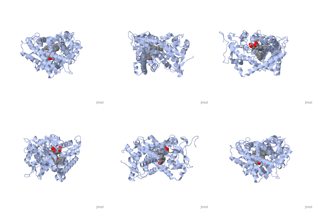
Now rotate the molecule by clicking and dragging in the window with your cursor or using the scroll wheel on your mouse. Re-size the molecule by holding down the shift key and dragging up and down. Rotate and re-size the molecule until you can clearly see that there are 2 molecules shown in a "space-filling" representation in the middle of the protein (they are almost perpendicular to each other, and almost touching). These are a heme molecule, which is absolutely vital for the enzyme's function, and a second molecule (alpha-naphthoflavone) which is a compound about to be metabolized.
As you go through this tutorial, rotate and re-size the molecules as necessary to see the concepts being illustrated. You might also find it useful to toggle the spin or quality of the display.
| |||||||||||

