Interacting with the Molecular Display
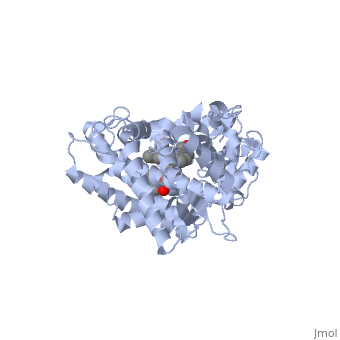
In this tutorial, the blue links are standard hyperlinks. The green links show you a particular view, or scene, of the molecule in the interactive window to the right. As you go through the text, click on the green links to show the structural features being highlighted. The first example illustrated here is the second protein discovered in the CYP 1 family, in subfamily A (generally referred to as CYP1A2). This protein is shown in an interactive window to the right, and comes from the PDB entry 2hi4.
Turn off/on (toggle) spinning of the protein by clicking on the button below the structure. The quality of the molecule image can also be increased by clicking the "toggle quality" button, although displaying it this way may decrease the smoothness when the molecule is rotating.
Now rotate the molecule by clicking and dragging in the window with your cursor or using the scroll wheel on your mouse. Re-size the molecule by holding down the shift key and dragging up and down. Rotate and re-size the molecule until you can clearly see that there are 2 molecules shown in a "space-filling" representation in the middle of the protein (they are almost perpendicular to each other, and almost touching). These are a heme molecule, which is absolutely vital for the enzyme's function, and a second molecule (𝛂-naphthoflavone) which is a compound about to be metabolized.
As you go through this tutorial, rotate and re-size the molecules as necessary to see the concepts being illustrated. You might also find it useful to toggle the spin or quality of the display.
The Heme Group is Vital for the Enzyme's Action
Let's look at the heme now. Click the following link to hide the rest of the protein and focus on the by itself. Carbon atoms are shown in grey, nitrogen in blue, oxygen in red, and the central iron atom in orange. The Iron atom is a vital center for oxidation of substrates (drugs or other xenobiotics). Rotate and re-size as necessary to shown that the heme is highly planar. Do you recognize the 4 pyrrole rings within the heme?
Examine the iron atom in the heme. It is capable of forming 6 bonds. Four of those are with the 4 pyrrole rings that make up the porphyrin ring system. The fifth and sixth bonds are made to atoms above and below the plane of heme ring. The fifth is made to a cysteine residue present on the protein. The final bond is made to an oxygen molecule (not shown). This molecular oxygen is activated to aid in oxidation of substrates and would appear directly between the substrate and the iron atom.
The cysteine residue that coordinates with the iron atom is vital; it helps to polarize the heme allowing heme to bind with oxygen. This residue is highly conserved throughout all of the known CYP450s. The bond between the sulfur atom of the cysteine residue and the heme ring is shown in yellow. Note that the sixth site for a bond with iron remains unfilled and available to bind to oxygen. Although the presence of the cysteine residue is invariable in all CYPs, the other residues that surround it may vary. This can affect the shape of the active site and the drugs that can bind there.
Molecules Can be Oxidized at More than One Position
Now let's take a look at being oxidized while noting its orientation relative to the heme group. Re-size and rotate the molecules until you can see how the two molecules are oriented in relationship to each other. The flavone is metabolized (oxidized) by introduction of a hydroxy group onto the phenyl ring attached to the tricyclic ring system. Note particularly the distance and orientation of the heme Iron and the phenyl substituent.
When some drugs are administered to humans, the metabolites observed may be the result of oxidation at more than one position in the molecule. What factors do you think might contribute to this? Several factors may come into play.
First, more than one CYP isoform may be contributing, with each isoform preferring different sites of attack.
Considering only one CYP, like that shown here, there is also the possibility that attack might occur at more than one position, giving more than one metabolite. What factors do you think might lead to this?
In this example, purely based on the distance between the phenyl ring of the flavone and the heme iron, oxidation might be expected to take place at the 4 (para) position. However, other factors may also help determine the regioselectivity of the metabolism (selectivity for oxidation at the different possible positions). One of these factors is the relative reactivity of the various positions on the substrate.
Different CYPs can be Selective for Different Drugs
Different CYPs have preferences for oxidizing different substrates. Some of them have a much narrower range of molecular structures that they can oxidize. CYP1A2 preferentially oxidizes molecules that are relatively large and planar, as in this structure [1]. The complementary size and shape of the drug and binding site makes them a good match for each other.
The substrate (flavone) shown here is different from many other substrates of this CYP. Besides being a substrate of CYP1A2, it is also a competitive inhibitor[2]. It can inhibit the metabolism of other CYP1A2 substrates because it binds very tightly to it. Thus, while the CYP is inhibited, it is unavailable to metabolize other substrates (e.g. drugs). Therefore, if a second drug that is a CYP1A2 substrate is co-administered with this compound, it might not be broken down as expected, and could build up to toxic levels.
The reason that this flavone is bound so tightly to the enzyme is that its shape and electronic charges are complementary to the binding pocket. This is examined next.
Amino Acids in the Binding Pocket Affect Drug Selectivity
First examine the shape of the around the flavone. The range of bluish-green to reddish-orange colored areas on the surface differentiate how close(red), or how far (blue), in proximity the flavone is to the binding pocket. These contacts can be caused by ionic, hydrophobic, or hydrogen bonding. Given the structure of the flavone, what forces do you suspect may be responsible for binding the enzyme so tightly?
In the next scene, the Van der Waals surface of the is displayed. The portions of the cavity involved in binding are shown as orange patches. These are a result of specific amino acid residues that form the surface of the binding pocket. Clicking on this will show the surface of the flavone and a few of the most important amino acid residues responsible for binding.
As you rotate the molecule, look at how each of the amino acid residues at the active site is interacting with parts of the flavone. Can you predict what kinds of interactions (H-bonds, lipophilic, or ionic) might be made? Examine the hydrophobic portions of the inhibitor. What kind of residues would interact with these? Hover the cursor over each residue and examine the label that appears (you may have to re-size the molecule to pick the individual atoms). Is the label consistent with your expectations for these kinds of interactions? Since the CYPs largely metabolize hydrophobic substrates, hydrophobic interactions are very important for binding many substrates. That is clearly the case here.
CYP450 3A4 and Its Malleable Active Site
The next ligand discussed fits exceptionally well into the binding pocket due to the principle of induced fit. Induced fit occurs when a drug binds to a protein and causes a conformational change that leads to tighter binding. Thus the "fit" of the drug to the protein is "induced" in many cases. A molecule that is able to induce a fit to a protein might be expected to exhibit competitive inhibition.
The CYP3A4 isoform, PDB entry 1tqn, is involved in metabolizing over 50% of marketed drugs, and accounts for approximately 40% of hepatic CYP enzymes. This helps to make sense of the fact that it is the enzyme most commonly associated with undesired drug-drug interactions. CYP3A4 differs from the CYP1A2 isoform considered above, as well as most other CYPs, because of the diversity of drugs it can metabolize. A number of these drugs are known to cause potentially dangerous or even fatal interactions. The reason that CYP3A4 is involved in the metabolism of so many drugs appears to be related to the size and flexibility of its binding pocket, which can accommodate a number of fairly large drugs. A few drugs considered to be "large" include macrolide antibiotic erythromycin, the anticancer drug taxol, the immunosuppressant cyclosporine, and several statins.
Examining the way drugs bind to CYP3A4 gives some insight into its selectivity and the way it may be influence the pharmacokinetics of the drug. In the crystal structure of CYP3A4 bound to erythromycin, the volume of the active site is notably different than the unbound protein. With erythromycin being a fairly large drug (MW 733.9), CYP450 isoforms with smaller binding pockets (e.g. CYP1A2) cannot accommodate it and therefore cannot metabolize it. Due to its flexible binding pocket, CYP3A4 can also accommodate a diversity of other, smaller substrates.
Effect of Drug Binding on a CYP450 Active Site
The following scene shows the ligand-free form of . The next scene shows the of CYP3A4 without a substrate. Examine the size and shape of the cavity by rotating and resizing the molecule. Observe that the cavity extends within a short distance of the heme ring. In order for a drug to be oxidized, it must come quite close to the oxygen atom held by the heme.
Rotate and re-size the molecule until you can clearly see that there are 2 regions open to the outside of the molecule. Note that the opening to the cavity does not seem large enough to allow a drug of any significant size to enter. In order for a larger drug to enter the inside of the enzyme, a change in its shape must occur.
Now, take a look at this scene showing (PDB entry 2j0d) with the surface of its binding pocket obviously altered. In this case, CYP3A4 has undergone a conformational change as a result of binding to erythromycin. Note the size and shape of the cavity, and the apparent size of the opening. In comparing the bound and unbound structures, it looks as though the enzyme has adopted a conformation that allows the drug to bind more tightly than its initial interaction. This, again, is induced fit.
Complex Drug Interactions with CYP450s
There are multiple types of interactions when considering CYP systems. Competitive inhibition follows fairly predictable rules. In this case, two drugs are competing for the same active site and one drug slows down the metabolism of the other. However, some drugs metabolized by CYP3A4 are known to have more complex interactions.
It has been demonstrated that two or more smaller molecules may bind within an active site at the same time. In this case, drug metabolism can, strangely enough, actually be increased. It has been proposed that when only one molecule of a smaller drug (let's call this Drug A) is bound to the active site, that the extent of Drug A 's metabolism can be minimal due to the relatively large cavity. One explanation may be that Drug A isn't held in a sufficient orientation to the heme Iron. For optimum metabolism, the heme should bond with an Oxygen of the ligand. However, when two molecules of a smaller drug bind at the same time, one molecule may help in forcing the other molecule to retain proper orientation; thus, improving catalytic efficiency. This theory of two drugs binding simultaneously to a CYP may influence its pharmacokinetics is illustrated by the following structure of ketoconazole bound to a CYP enzyme (2v0m) [3].
Ketoconazole is an anti-fungal drug that can have unusual pharmacokinetics; its apparent plasma concentration does not reflect what we would traditionally expect when considering the dose given. In the structure shown next, are bound to the CYP. You can see this interaction better if we . One of the ketoconazole molecules is bound directly to the heme ring, while the second molecule has taken up residence in the pocket and appears to be ensuring the first one remain in place. The unusual pharmacokinetics of ketoconazole may be explained by the fact that as its plasma concentration increases, the activity of the enzyme is altered due to two drugs now being bound.
Irreversible inhibition of CYP450s
When we examined CYP1A2 above, the flavone inhibited the enzyme simply by virtue of having such complementary features to the binding site. It binds so tightly that it physically prevents other drugs from binding to it. Another way that a drug can inhibit CYP450 enzymes is by formation of a covalent bond that deactivates the active site. This happens with a well-known inhibitor of CYP3A4, . Ritonavir is a HIV protease inhibitor routinely prescribed in combination with other antivirals. Its efficacy as part of a drug "cocktail" stems from the fact that it is a potent "irreversible inhibitor" of CYP3A4. Irreversible inhibition differs from most cases of competitive inhibition in that the enzyme is permanently deactivated, and must be re-synthesized by the cell.
In this case, we have taken advantage of the inhibition of CYP3A4 to prevent it from metabolizing the other antivirals of which ritonavir is co-administered. As you may expect though, extreme caution must be taken to prevent toxicity when other medications are taken concurrently. This is relatively easy to control when only one pharmacy is dispensing all of the medications a patient is prescribed. When more than one pharmacy is involved, however, serious interactions may be overlooked due to one pharmacy being unaware of the medications a patient is receiving from the other pharmacy.
Examine the orientation of ritonavir relative to the heme group. Now let's look at the . Notice the complementary shape of the drug to the cavity. In contrast to erythromycin, there has been a covalent bond formed between the of the drug and the heme ring. Do you see how the thiazole has bound to the heme?
In the next section below, we will examine another factor that causes one drug to be metabolized by one CYP, while another might be metabolized by a second one.