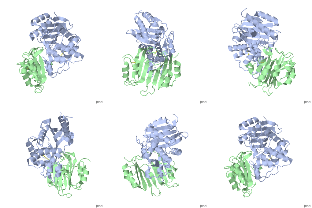
2g2u
From Proteopedia
| Line 1: | Line 1: | ||
| - | [[Image:2g2u.gif|left|200px]] | + | [[Image:2g2u.gif|left|200px]] |
| - | + | ||
| - | '''Crystal Structure of the SHV-1 Beta-lactamase/Beta-lactamase inhibitor protein (BLIP) complex''' | + | {{Structure |
| + | |PDB= 2g2u |SIZE=350|CAPTION= <scene name='initialview01'>2g2u</scene>, resolution 1.60Å | ||
| + | |SITE= | ||
| + | |LIGAND= | ||
| + | |ACTIVITY= [http://en.wikipedia.org/wiki/Beta-lactamase Beta-lactamase], with EC number [http://www.brenda-enzymes.info/php/result_flat.php4?ecno=3.5.2.6 3.5.2.6] | ||
| + | |GENE= bla, shv1 ([http://www.ncbi.nlm.nih.gov/Taxonomy/Browser/wwwtax.cgi?mode=Info&srchmode=5&id=573 Klebsiella pneumoniae]) | ||
| + | }} | ||
| + | |||
| + | '''Crystal Structure of the SHV-1 Beta-lactamase/Beta-lactamase inhibitor protein (BLIP) complex''' | ||
| + | |||
==Overview== | ==Overview== | ||
| Line 7: | Line 16: | ||
==About this Structure== | ==About this Structure== | ||
| - | 2G2U is a [ | + | 2G2U is a [[Protein complex]] structure of sequences from [http://en.wikipedia.org/wiki/Klebsiella_pneumoniae Klebsiella pneumoniae] and [http://en.wikipedia.org/wiki/Streptomyces_clavuligerus Streptomyces clavuligerus]. Full crystallographic information is available from [http://oca.weizmann.ac.il/oca-bin/ocashort?id=2G2U OCA]. |
==Reference== | ==Reference== | ||
| - | Structural and computational characterization of the SHV-1 beta-lactamase-beta-lactamase inhibitor protein interface., Reynolds KA, Thomson JM, Corbett KD, Bethel CR, Berger JM, Kirsch JF, Bonomo RA, Handel TM, J Biol Chem. 2006 Sep 8;281(36):26745-53. Epub 2006 Jun 29. PMID:[http:// | + | Structural and computational characterization of the SHV-1 beta-lactamase-beta-lactamase inhibitor protein interface., Reynolds KA, Thomson JM, Corbett KD, Bethel CR, Berger JM, Kirsch JF, Bonomo RA, Handel TM, J Biol Chem. 2006 Sep 8;281(36):26745-53. Epub 2006 Jun 29. PMID:[http://www.ncbi.nlm.nih.gov/pubmed/16809340 16809340] |
[[Category: Beta-lactamase]] | [[Category: Beta-lactamase]] | ||
[[Category: Klebsiella pneumoniae]] | [[Category: Klebsiella pneumoniae]] | ||
| Line 29: | Line 38: | ||
[[Category: shv-1]] | [[Category: shv-1]] | ||
| - | ''Page seeded by [http://oca.weizmann.ac.il/oca OCA ] on Thu | + | ''Page seeded by [http://oca.weizmann.ac.il/oca OCA ] on Thu Mar 20 16:59:43 2008'' |
Revision as of 14:59, 20 March 2008
| |||||||
| , resolution 1.60Å | |||||||
|---|---|---|---|---|---|---|---|
| Gene: | bla, shv1 (Klebsiella pneumoniae) | ||||||
| Activity: | Beta-lactamase, with EC number 3.5.2.6 | ||||||
| Coordinates: | save as pdb, mmCIF, xml | ||||||
Crystal Structure of the SHV-1 Beta-lactamase/Beta-lactamase inhibitor protein (BLIP) complex
Overview
Beta-lactamase inhibitor protein (BLIP) binds a variety of class A beta-lactamases with affinities ranging from micromolar to picomolar. Whereas the TEM-1 and SHV-1 beta-lactamases are almost structurally identical, BLIP binds TEM-1 approximately 1000-fold tighter than SHV-1. Determining the underlying source of this affinity difference is important for understanding the molecular basis of beta-lactamase inhibition and mechanisms of protein-protein interface specificity and affinity. Here we present the 1.6A resolution crystal structure of SHV-1.BLIP. In addition, a point mutation was identified, SHV D104E, that increases SHV.BLIP binding affinity from micromolar to nanomolar. Comparison of the SHV-1.BLIP structure with the published TEM-1.BLIP structure suggests that the increased volume of Glu-104 stabilizes a key binding loop in the interface. Solution of the 1.8A SHV D104K.BLIP crystal structure identifies a novel conformation in which this binding loop is removed from the interface. Using these structural data, we evaluated the ability of EGAD, a program developed for computational protein design, to calculate changes in the stability of mutant beta-lactamase.BLIP complexes. Changes in binding affinity were calculated within an error of 1.6 kcal/mol of the experimental values for 112 mutations at the TEM-1.BLIP interface and within an error of 2.2 kcal/mol for 24 mutations at the SHV-1.BLIP interface. The reasonable success of EGAD in predicting changes in interface stability is a promising step toward understanding the stability of the beta-lactamase.BLIP complexes and computationally assisted design of tight binding BLIP variants.
About this Structure
2G2U is a Protein complex structure of sequences from Klebsiella pneumoniae and Streptomyces clavuligerus. Full crystallographic information is available from OCA.
Reference
Structural and computational characterization of the SHV-1 beta-lactamase-beta-lactamase inhibitor protein interface., Reynolds KA, Thomson JM, Corbett KD, Bethel CR, Berger JM, Kirsch JF, Bonomo RA, Handel TM, J Biol Chem. 2006 Sep 8;281(36):26745-53. Epub 2006 Jun 29. PMID:16809340
Page seeded by OCA on Thu Mar 20 16:59:43 2008

