We apologize for Proteopedia being slow to respond. For the past two years, a new implementation of Proteopedia has been being built. Soon, it will replace this 18-year old system. All existing content will be moved to the new system at a date that will be announced here.
Sandbox Reserved 951
From Proteopedia
(Difference between revisions)
| Line 36: | Line 36: | ||
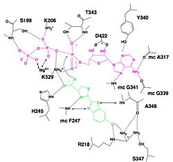
[[Image:Luciferin_bounding_to_luciferase.jpg|250px|right|thumb|Hydrogen bonding between Luciferase and substrates luciferin (green), ATP (violet) and Mg2+,<ref name=''first''>[http://www.photobiology.info/ Photobiology]</ref>]] | [[Image:Luciferin_bounding_to_luciferase.jpg|250px|right|thumb|Hydrogen bonding between Luciferase and substrates luciferin (green), ATP (violet) and Mg2+,<ref name=''first''>[http://www.photobiology.info/ Photobiology]</ref>]] | ||
=====Interaction with ATP===== | =====Interaction with ATP===== | ||
| - | We find a signal motif in luciferase which is <scene name='60/604470/Atp_binding_signal_motif/1'>[STG]-[STG]-G-[ST]-[ST]-[TSE]-[GS]-x-[PALIVM]-K</scene> | + | We find a signal motif in luciferase which is <scene name='60/604470/Atp_binding_signal_motif/1'>[STG]-[STG]-G-[ST]-[ST]-[TSE]-[GS]-x-[PALIVM]-K</scene>. Here some residues like lysine are always conserved. This pattern enables ATP binding thanks to hydrogen bonds between residues and phosphates of ATP. There is another pattern : <scene name='60/604470/Adenosine_ring_binding/1'>[YFW]-[GASW]-x-[TSA]-E</scene> which takes a particular conformation because of hydrogen bonds between residues and maintain the adenosin ring of ATP.<ref name =''fifth''>PMID:8805533</ref>, <ref name=''second''>[http://www.photobiology.info/ Photobiology]</ref> |
=====Interaction with luciferin===== | =====Interaction with luciferin===== | ||
Luciferase holds the luciferin with the specific residues <scene name='60/604470/Residues_helding_luciferin/1'>arginin 218, phenylalanin 247, serin 347 and adenin 348</scene>, still with hydrogen bounds. Those bindings make the carboxylate oxygen of luciferin points toward the α phosphate of ATP, so the oxygen is well-positionned to attack the α phosphate. This promotes the luciferin-AMP formation.<ref name =''sixth''>PMID:8805533</ref>, <ref name=''third''>[http://www.photobiology.info/ Photobiology]</ref> | Luciferase holds the luciferin with the specific residues <scene name='60/604470/Residues_helding_luciferin/1'>arginin 218, phenylalanin 247, serin 347 and adenin 348</scene>, still with hydrogen bounds. Those bindings make the carboxylate oxygen of luciferin points toward the α phosphate of ATP, so the oxygen is well-positionned to attack the α phosphate. This promotes the luciferin-AMP formation.<ref name =''sixth''>PMID:8805533</ref>, <ref name=''third''>[http://www.photobiology.info/ Photobiology]</ref> | ||
Revision as of 14:19, 9 January 2015
| |||||||||||
References
- ↑ Welsh DK, Kay SA. Bioluminescence imaging in living organisms. Curr Opin Biotechnol. 2005 Feb;16(1):73-8. PMID:15722018 doi:http://dx.doi.org/10.1016/j.copbio.2004.12.006
- ↑ 2.0 2.1 2.2 2.3 2.4 2.5 2.6 Conti E, Franks NP, Brick P. Crystal structure of firefly luciferase throws light on a superfamily of adenylate-forming enzymes. Structure. 1996 Mar 15;4(3):287-98. PMID:8805533
- ↑ Marques SM, Esteves da Silva JC. Firefly bioluminescence: a mechanistic approach of luciferase catalyzed reactions. IUBMB Life. 2009 Jan;61(1):6-17. PMID:18949818 doi:10.1002/iub.134
- ↑ Photobiology
- ↑ Photobiology
- ↑ Photobiology
- ↑ Marques SM, Esteves da Silva JC. Firefly bioluminescence: a mechanistic approach of luciferase catalyzed reactions. IUBMB Life. 2009 Jan;61(1):6-17. PMID:18949818 doi:10.1002/iub.134
- ↑ Hosseinkhani S. Molecular enigma of multicolor bioluminescence of firefly luciferase. Cell Mol Life Sci. 2011 Apr;68(7):1167-82. doi: 10.1007/s00018-010-0607-0. Epub, 2010 Dec 28. PMID:21188462 doi:http://dx.doi.org/10.1007/s00018-010-0607-0
- ↑ Inouye S. Firefly luciferase: an adenylate-forming enzyme for multicatalytic functions. Cell Mol Life Sci. 2010 Feb;67(3):387-404. Epub 2009 Oct 27. PMID:19859663 doi:10.1007/s00018-009-0170-8


