We apologize for Proteopedia being slow to respond. For the past two years, a new implementation of Proteopedia has been being built. Soon, it will replace this 18-year old system. All existing content will be moved to the new system at a date that will be announced here.
Tachyplesin
From Proteopedia
(Difference between revisions)
| Line 43: | Line 43: | ||
== Mode of action == | == Mode of action == | ||
| - | TP-I has high affinity to negatively charged cell membrane containing LPS and also has ability to permeabilize the cell membrane of pathogens. Docking model suggests strong interaction between cationic residues of TP-I with phosphate group and saccharides of LPS. Also, interaction between hydrophobic residues of TP-I with acyl chains of LPS was observed which strengthens the TP-I/LPS interaction<ref name=Hong>Hong, Jun, et al. "Mechanism of Tachyplesin I injury to bacterial membranes and intracellular enzymes, determined by laser confocal scanning microscopy and flow cytometry." Microbiological research (2014). Ultimately, binding of TP-I/LPS neutralizes LPS, which is widely considered as endotoxin, and disrupts membrane function. | + | TP-I has high affinity to negatively charged cell membrane containing LPS and also has ability to permeabilize the cell membrane of pathogens. Docking model suggests strong interaction between cationic residues of TP-I with phosphate group and saccharides of LPS. Also, interaction between hydrophobic residues of TP-I with acyl chains of LPS was observed which strengthens the TP-I/LPS interaction<ref name=Hong>Hong, Jun, et al. "Mechanism of Tachyplesin I injury to bacterial membranes and intracellular enzymes, determined by laser confocal scanning microscopy and flow cytometry." Microbiological research (2014)</ref>. Ultimately, binding of TP-I/LPS neutralizes LPS, which is widely considered as endotoxin, and disrupts membrane function. |
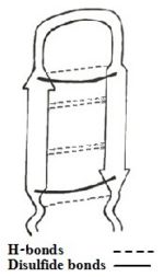
In addition to LPS binding, footpriting analysis has revealed the binding of TP-I to DNA by interacting specifically in minor groove of DNA duplex. The interaction between TP-I and DNA is contributed by secondary structure of the peptide which contains an antiparallel beta-sheet constrained by two disulfide bridges and connected by β-turn <ref name=Yonezawa>PMID:1372516</ref>. | In addition to LPS binding, footpriting analysis has revealed the binding of TP-I to DNA by interacting specifically in minor groove of DNA duplex. The interaction between TP-I and DNA is contributed by secondary structure of the peptide which contains an antiparallel beta-sheet constrained by two disulfide bridges and connected by β-turn <ref name=Yonezawa>PMID:1372516</ref>. | ||
By binding to DNA and RNA TP-I inhibits the synthesis of macromolecules. | By binding to DNA and RNA TP-I inhibits the synthesis of macromolecules. | ||
| - | In summary, three processes might happen upon TP-I exposure: (1) Bacterial cell membranes are penetrated without disruption of the membrane and the peptide reaches the inner structures of the cell, damaging critical intracellular targets and interfering with intracellular functions and normal metabolism. (2) Pores are formed in the cell wall, causing leakage of intracellular content, leading to cell death. (3) DNA, RNA or protein synthesis are inhibited, killing the bacteria.<ref name=Hong>Hong, Jun, et al. "Mechanism of tachyplesin I injury to bacterial membranes and intracellular enzymes, determined by laser confocal scanning microscopy and flow cytometry." Microbiological research (2014) | + | In summary, three processes might happen upon TP-I exposure: (1) Bacterial cell membranes are penetrated without disruption of the membrane and the peptide reaches the inner structures of the cell, damaging critical intracellular targets and interfering with intracellular functions and normal metabolism. (2) Pores are formed in the cell wall, causing leakage of intracellular content, leading to cell death. (3) DNA, RNA or protein synthesis are inhibited, killing the bacteria.<ref name=Hong>Hong, Jun, et al. "Mechanism of tachyplesin I injury to bacterial membranes and intracellular enzymes, determined by laser confocal scanning microscopy and flow cytometry." Microbiological research (2014)</ref>. |
== Importance and relevance == | == Importance and relevance == | ||
Revision as of 17:17, 23 January 2015
| |||||||||||
References
- ↑ 1.0 1.1 1.2 1.3 Laederach A, Andreotti AH, Fulton DB. Solution and micelle-bound structures of tachyplesin I and its active aromatic linear derivatives. Biochemistry. 2002 Oct 15;41(41):12359-68. PMID:12369825
- ↑ 2.0 2.1 Chen, Yixin, et al. "RGD-Tachyplesin inhibits tumor growth." Cancer research 61.6 (2001): 2434-2438.
- ↑ 3.0 3.1 Saravanan R, Mohanram H, Joshi M, Domadia PN, Torres J, Ruedl C, Bhattacharjya S. Structure, activity and interactions of the cysteine deleted analog of tachyplesin-1 with lipopolysaccharide micelle: Mechanistic insights into outer-membrane permeabilization and endotoxin neutralization. Biochim Biophys Acta. 2012 Mar 23;1818(7):1613-1624. PMID:22464970 doi:10.1016/j.bbamem.2012.03.015
- ↑ Nakamura, Takanori, et al. "Tachyplesin, a class of antimicrobial peptide from the hemocytes of the horseshoe crab (Tachypleus tridentatus). Isolation and chemical structure." Journal of Biological Chemistry 263.32 (1988): 16709-16713
- ↑ 5.0 5.1 Kushibiki T, Kamiya M, Aizawa T, Kumaki Y, Kikukawa T, Mizuguchi M, Demura M, Kawabata SI, Kawano K. Interaction between tachyplesin I, an antimicrobial peptide derived from horseshoe crab, and lipopolysaccharide. Biochim Biophys Acta. 2014 Jan 2;1844(3):527-534. doi:, 10.1016/j.bbapap.2013.12.017. PMID:24389234 doi:http://dx.doi.org/10.1016/j.bbapap.2013.12.017
- ↑ 6.0 6.1 6.2 Hong, Jun, et al. "Mechanism of Tachyplesin I injury to bacterial membranes and intracellular enzymes, determined by laser confocal scanning microscopy and flow cytometry." Microbiological research (2014)
- ↑ Yonezawa A, Kuwahara J, Fujii N, Sugiura Y. Binding of tachyplesin I to DNA revealed by footprinting analysis: significant contribution of secondary structure to DNA binding and implication for biological action. Biochemistry. 1992 Mar 24;31(11):2998-3004. PMID:1372516
- ↑ Lipsky A, Cohen A, Ion A, Yedidia I. Genetic transformation of Ornithogalum via particle bombardment and generation of Pectobacterium carotovorum-resistant plants. Plant Sci. 2014 Nov;228:150-8. doi: 10.1016/j.plantsci.2014.02.002. Epub 2014 Feb, 12. PMID:25438795 doi:http://dx.doi.org/10.1016/j.plantsci.2014.02.002
Proteopedia Page Contributors and Editors (what is this?)
Shulamit Idzikowski, Janak Raj Joshi, Michal Harel, Alexander Berchansky, Joel L. Sussman, Angel Herraez, Jaime Prilusky


