This old version of Proteopedia is provided for student assignments while the new version is undergoing repairs. Content and edits done in this old version of Proteopedia after March 1, 2026 will eventually be lost when it is retired in about June of 2026.
Apply for new accounts at the new Proteopedia. Your logins will work in both the old and new versions.
Ubc9
From Proteopedia
(Difference between revisions)
| Line 1: | Line 1: | ||
| - | + | ==Your Heading Here (maybe something like 'Structure')== | |
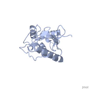
| + | <StructureSection load='1U9A' size='350' side='right' caption='Human Ubiquitin Conjugating Protein ''Ubc9'' ' scene=''> | ||
== Structure == | == Structure == | ||
| - | ''Ubc9'' exhibits a single domain structure consisting of both alpha helices and beta-pleated sheets. | + | ''Ubc9'' exhibits a single domain structure consisting of both alpha helices and beta-pleated sheets. Cys93 has been identified as the active residue and is located on a non-secondary structured loop of amino acids (78-108) between beta sheet four and alpha helix two ^1^ . |
== Function == | == Function == | ||
| Line 20: | Line 21: | ||
- | - | ||
| + | |||
| + | Anything in this section will appear adjacent to the 3D structure and will be scrollable. | ||
| + | |||
| + | </StructureSection> | ||
| + | |||
== References == | == References == | ||
1. Reverter, D., & Lima, C. D. (2005). Insights into E3 ligase activity revealed by a SUMO-RanGAP1-Ubc9-Nup358 complex. Nature, 435(June), 687–692. doi:10.1038/nature03588 | 1. Reverter, D., & Lima, C. D. (2005). Insights into E3 ligase activity revealed by a SUMO-RanGAP1-Ubc9-Nup358 complex. Nature, 435(June), 687–692. doi:10.1038/nature03588 | ||
<references/> | <references/> | ||
Revision as of 15:21, 23 February 2015
Your Heading Here (maybe something like 'Structure')
| |||||||||||
References
1. Reverter, D., & Lima, C. D. (2005). Insights into E3 ligase activity revealed by a SUMO-RanGAP1-Ubc9-Nup358 complex. Nature, 435(June), 687–692. doi:10.1038/nature03588
