Myoglobin
From Proteopedia
| Line 9: | Line 9: | ||
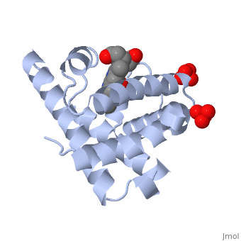
<StructureSection load='1a6m' size='350' side='right' caption='Structure of Sperm whale myoglobin with O2 and sulfate (PDB entry [[1a6m]])' scene=''> | <StructureSection load='1a6m' size='350' side='right' caption='Structure of Sperm whale myoglobin with O2 and sulfate (PDB entry [[1a6m]])' scene=''> | ||
| - | [[Myoglobin]] is a globular protein whose function is to store molecular oxygen. The fold of the protein is conserved but the sequence is more | + | [[Myoglobin]] is a globular protein whose function is to store molecular oxygen. The fold of the protein is conserved but the sequence is more <scene name='23/238129/Conserved_cartoon/1'>variable</scene>. {{Template:ColorKey_ConSurf}} |
The globin consists mostly of [[Helices in Proteins|alpha helices]] shown in <scene name='23/238129/2ndary_structure/2'>pink</scene>; it has no beta sheets and its nonhelical segments mostly serve as links that connect the helices. Look down the barrel of some of the longer helices. Are they all straight? The eight structurally conserved alpha helices are labelled <scene name='23/238129/Helix_labels/1'>A through H</scene>. The protein is colored as a N-->C rainbow in this view; the N terminus is blue, while the C terminus is red. | The globin consists mostly of [[Helices in Proteins|alpha helices]] shown in <scene name='23/238129/2ndary_structure/2'>pink</scene>; it has no beta sheets and its nonhelical segments mostly serve as links that connect the helices. Look down the barrel of some of the longer helices. Are they all straight? The eight structurally conserved alpha helices are labelled <scene name='23/238129/Helix_labels/1'>A through H</scene>. The protein is colored as a N-->C rainbow in this view; the N terminus is blue, while the C terminus is red. | ||
Revision as of 03:53, 29 March 2015
|
Caution: The text in this article and has not been updated to reflect what is actually available on this page. There is no zoom slider and no animate button. These were formerly present when an earlier version of Proteopedia supported Kinemages. A volunteer is needed to clean up and improve this article on a pedagogically and historically important protein. Green links are needed! Text referring to the now absent Kinemage is in gray. Eric Martz 01:09, 13 September 2014 (IDT) |
| |||||||||||

</font>
Former exercise in large part by John H. Connor (present address: Department of Microbiology, Boston University School of Medicine, 850 Harrison Ave, Boston, MA, 02118, USA). Revised by Ann Taylor
3D Structures of Myoglobin
Updated on 29-March-2015 Myoglobin (Mb) is an oxygen binding protein found in muscle tissue. It contains a heme group. Metmyoglobin (MMb) is the oxidized form of myoglobin.
((3qm5, 3qm6 – btMb – blackfin tuna
External Resources
Proteopedia Page Contributors and Editors (what is this?)
Michal Harel, Ann Taylor, Alexander Berchansky, Joel L. Sussman, Eric Martz, Jaime Prilusky, Karsten Theis, Karl Oberholser, Eran Hodis, Judy Voet, David Canner
