We apologize for Proteopedia being slow to respond. For the past two years, a new implementation of Proteopedia has been being built. Soon, it will replace this 18-year old system. All existing content will be moved to the new system at a date that will be announced here.
Sandbox Reserved 1063
From Proteopedia
(Difference between revisions)
| Line 11: | Line 11: | ||
== Structural Highlights == | == Structural Highlights == | ||
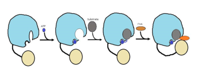
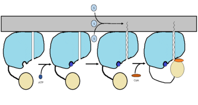
| - | FadD13 is an ACSVL enzyme that can accept lipids up to 26 carbons as well as being a [http://en.wikipedia.org/wiki/Peripheral_membrane_protein peripheral-membrane protein]<ref>PMID: 20027301</ref>. Unlike other ACSVL proteins, FadD13 is soluble. There are numerous aspects of its structure that affects the way this protein functions. The <scene name='69/694230/Atp_and_amp_binding_region/13'>ATP/AMP binding region</scene> is comprised of two separate motifs. The first motif is composed of residues 164-TSGTTGHPKG173-173 shown in red which binds to the phosphate group. The second motif is comprised of residues 298-VQGYALTE-305 shown in blue which binds to the adenine group<ref>DOI: 10.1371/journal.pone.0008387</ref>. Upon binding of ATP or AMP, FadD13 is activated. There are two domains of this protein, a larger N-terminal domain (<scene name='69/694230/N-terminal_domain/5'>residues 1-395</scene>) shown in blue and a smaller C-terminal domain (<scene name='69/694230/C-terminal_domain/1'>residues 402-503</scene>). These domains are held together by a six amino acid linker (<scene name='69/694230/Residues_396-401/1'>residues 396-401</scene>) shown in purple. Inside the larger N-terminal domain is a hydrophobic tunnel, which allows large lipids/fatty acids, up to 26 carbons, to bind. The tunnel is capped by an arginine-rich lid loop that is involved in the peripheral binding of the enzyme to the membrane. | + | FadD13 is an ACSVL enzyme that can accept lipids up to 26 carbons as well as being a [http://en.wikipedia.org/wiki/Peripheral_membrane_protein peripheral-membrane protein]<ref>PMID: 20027301</ref>. Unlike other ACSVL proteins, FadD13 is soluble. There are numerous aspects of its structure that affects the way this protein functions. The <scene name='69/694230/Atp_and_amp_binding_region/13'>ATP/AMP binding region</scene> is comprised of two separate motifs. The first motif is composed of residues 164-TSGTTGHPKG173-173 shown in red which binds to the phosphate group. The second motif is comprised of residues 298-VQGYALTE-305 shown in blue which binds to the adenine group<ref>DOI: 10.1371/journal.pone.0008387</ref>. Upon binding of ATP or AMP, FadD13 is activated. There are two domains of this protein, a larger N-terminal domain (<scene name='69/694230/N-terminal_domain/5'>residues 1-395</scene>) shown in blue and a smaller C-terminal domain (<scene name='69/694230/C-terminal_domain/1'>residues 402-503</scene>). These domains are held together by a six amino acid linker (<scene name='69/694230/Residues_396-401/1'>residues 396-401</scene>) shown in purple. Inside the larger N-terminal domain is a hydrophobic tunnel, which allows large lipids/fatty acids, up to 26 carbons, to bind. The tunnel is capped by an arginine-rich lid loop that is involved in the peripheral binding of the enzyme to the membrane. Six key arginine residues,<scene name='69/694230/Arginine_surface_patch/3'>ARG 9, 17, 195, 197, 199, 244</scene> PMID: 22560731</ref> all play an important role for the enzyme to be able to bind to the cell membrane. |
== Function == | == Function == | ||
Revision as of 01:32, 21 April 2015
FadD13
| |||||||||||
References
- ↑ Watkins PA, Maiguel D, Jia Z, Pevsner J. Evidence for 26 distinct acyl-coenzyme A synthetase genes in the human genome. J Lipid Res. 2007 Dec;48(12):2736-50. Epub 2007 Aug 30. PMID:17762044 doi:http://dx.doi.org/M700378-JLR200
- ↑ Kochan G, Pilka ES, von Delft F, Oppermann U, Yue WW. Structural snapshots for the conformation-dependent catalysis by human medium-chain acyl-coenzyme A synthetase ACSM2A. J Mol Biol. 2009 May 22;388(5):997-1008. Epub 2009 Apr 1. PMID:19345228 doi:10.1016/j.jmb.2009.03.064
- ↑ Khare G, Gupta V, Gupta RK, Gupta R, Bhat R, Tyagi AK. Dissecting the role of critical residues and substrate preference of a Fatty Acyl-CoA Synthetase (FadD13) of Mycobacterium tuberculosis. PLoS One. 2009 Dec 21;4(12):e8387. doi: 10.1371/journal.pone.0008387. PMID:20027301 doi:10.1371/journal.pone.0008387
- ↑ Khare G, Gupta V, Gupta RK, Gupta R, Bhat R, Tyagi AK. Dissecting the role of critical residues and substrate preference of a Fatty Acyl-CoA Synthetase (FadD13) of Mycobacterium tuberculosis. PLoS One. 2009 Dec 21;4(12):e8387. doi: 10.1371/journal.pone.0008387. PMID:20027301 doi:10.1371/journal.pone.0008387
- ↑ Andersson CS, Lundgren CA, Magnusdottir A, Ge C, Wieslander A, Molina DM, Hogbom M. The Mycobacterium tuberculosis Very-Long-Chain Fatty Acyl-CoA Synthetase: Structural Basis for Housing Lipid Substrates Longer than the Enzyme. Structure. 2012 May 2. PMID:22560731 doi:10.1016/j.str.2012.03.012


