We apologize for Proteopedia being slow to respond. For the past two years, a new implementation of Proteopedia has been being built. Soon, it will replace this 18-year old system. All existing content will be moved to the new system at a date that will be announced here.
Sandbox Reserved 1063
From Proteopedia
(Difference between revisions)
| Line 10: | Line 10: | ||
== Structural Highlights == | == Structural Highlights == | ||
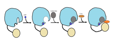
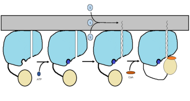
| - | FadD13 is an ACSVL enzyme that can accept lipids up to 26 carbons as well as being a [http://en.wikipedia.org/wiki/Peripheral_membrane_protein peripheral-membrane protein]<ref>PMID: 20027301</ref>. Unlike other ACSVL proteins, FadD13 is soluble. There are numerous aspects of its structure that affects the way this protein functions. The <scene name='69/694230/Fadd13_subunits/12'>ATP/AMP binding region</scene> is comprised of two separate motifs. The first motif is composed of residues 164-TSGTTGHPKG173-173 shown in red which binds to the phosphate group. The second motif is comprised of residues 298-VQGYALTE-305 shown in blue which binds to the adenine group<ref>DOI: 10.1371/journal.pone.0008387</ref>. Upon binding of ATP or AMP, FadD13 is activated. | + | FadD13 is composed of 503 amino acids which are divided into two domains. The larger of the two domains is the N-terminal domain composed of <scene name='69/694230/Fadd13_subunits/8'>residues 1-395</scene> shown in blue. The smaller of the two domains is the C-terminal domain composed of <scene name='69/694230/Fadd13_subunits/10'>residues 402-503</scene> shown in yellow. These two domains are held together by a flexible 6 amino acid linker (<scene name='69/694230/Fadd13_subunits/11'>residues 396-401</scene>) shown in black |
| + | ===Active Site=== | ||
| + | ===Surface Patch=== | ||
| + | ===Hydrophobic Tunnel=== | ||
| + | FadD13 is an ACSVL enzyme that can accept lipids up to 26 carbons as well as being a [http://en.wikipedia.org/wiki/Peripheral_membrane_protein peripheral-membrane protein]<ref>PMID: 20027301</ref>. Unlike other ACSVL proteins, FadD13 is soluble. There are numerous aspects of its structure that affects the way this protein functions. The <scene name='69/694230/Fadd13_subunits/12'>ATP/AMP binding region</scene> is comprised of two separate motifs. The first motif is composed of residues 164-TSGTTGHPKG173-173 shown in red which binds to the phosphate group. The second motif is comprised of residues 298-VQGYALTE-305 shown in blue which binds to the adenine group<ref>DOI: 10.1371/journal.pone.0008387</ref>. Upon binding of ATP or AMP, FadD13 is activated. Inside the larger N-terminal domain is a <scene name='69/694230/Fadd13_subunits/13'>hydrophobic tunnel</scene> composed of six beta sheets (beta 9-14) shown in green and two alpha helices (alpha 8-9) shown in red.The hydrophobic tunnel allows large lipids/fatty acids, up to 26 carbons, to bind. The tunnel is capped by an arginine and aromatic rich <scene name='69/694230/Fadd13_subunits/14'>lid loop</scene> shown in yellow that is involved in the peripheral binding of the enzyme to the membrane. Six key arginine residues, <scene name='69/694230/Fadd13_subunits/15'>Arg 9, 17, 195, 197, 199, and 244</scene> create a positively charged surface that is likely involved in initially recruiting FadD13 to the membrane. When these residues were replaced with hyrdrophobic alanine residues, membrane binding increased. This points to the important role of hydrophobic interactions in keeping the protein bound at the membrane<ref>PMID: 22560731</ref>. | ||
== Function == | == Function == | ||
Revision as of 15:41, 21 April 2015
FadD13
| |||||||||||
References
- ↑ Watkins PA, Maiguel D, Jia Z, Pevsner J. Evidence for 26 distinct acyl-coenzyme A synthetase genes in the human genome. J Lipid Res. 2007 Dec;48(12):2736-50. Epub 2007 Aug 30. PMID:17762044 doi:http://dx.doi.org/M700378-JLR200
- ↑ Kochan G, Pilka ES, von Delft F, Oppermann U, Yue WW. Structural snapshots for the conformation-dependent catalysis by human medium-chain acyl-coenzyme A synthetase ACSM2A. J Mol Biol. 2009 May 22;388(5):997-1008. Epub 2009 Apr 1. PMID:19345228 doi:10.1016/j.jmb.2009.03.064
- ↑ Khare G, Gupta V, Gupta RK, Gupta R, Bhat R, Tyagi AK. Dissecting the role of critical residues and substrate preference of a Fatty Acyl-CoA Synthetase (FadD13) of Mycobacterium tuberculosis. PLoS One. 2009 Dec 21;4(12):e8387. doi: 10.1371/journal.pone.0008387. PMID:20027301 doi:10.1371/journal.pone.0008387
- ↑ Khare G, Gupta V, Gupta RK, Gupta R, Bhat R, Tyagi AK. Dissecting the role of critical residues and substrate preference of a Fatty Acyl-CoA Synthetase (FadD13) of Mycobacterium tuberculosis. PLoS One. 2009 Dec 21;4(12):e8387. doi: 10.1371/journal.pone.0008387. PMID:20027301 doi:10.1371/journal.pone.0008387
- ↑ Andersson CS, Lundgren CA, Magnusdottir A, Ge C, Wieslander A, Molina DM, Hogbom M. The Mycobacterium tuberculosis Very-Long-Chain Fatty Acyl-CoA Synthetase: Structural Basis for Housing Lipid Substrates Longer than the Enzyme. Structure. 2012 May 2. PMID:22560731 doi:10.1016/j.str.2012.03.012
- ↑ Andersson CS, Lundgren CA, Magnusdottir A, Ge C, Wieslander A, Molina DM, Hogbom M. The Mycobacterium tuberculosis Very-Long-Chain Fatty Acyl-CoA Synthetase: Structural Basis for Housing Lipid Substrates Longer than the Enzyme. Structure. 2012 May 2. PMID:22560731 doi:10.1016/j.str.2012.03.012


