Regulator of G protein signaling
From Proteopedia
(Difference between revisions)
| Line 3: | Line 3: | ||
== RGS proteins == | == RGS proteins == | ||
Regulator of G-proteins signaling (RGS) proteins play a critical role in many G protein-dependent signaling pathways. Thus, RGS proteins have been implicated in a wide range of pathologies, including cancer, hypertension, arrhythmias, drug abuse and schizophrenia. RGS proteins ‘turn off’ heterotrimeric (αβγ) G-proteins and thereby determine the duration of G protein–mediated signaling events. Therefore, RGS proteins function as GTPase Activating Proteins (GAPs), and this GAP activity is mediated by allosteric interactions. | Regulator of G-proteins signaling (RGS) proteins play a critical role in many G protein-dependent signaling pathways. Thus, RGS proteins have been implicated in a wide range of pathologies, including cancer, hypertension, arrhythmias, drug abuse and schizophrenia. RGS proteins ‘turn off’ heterotrimeric (αβγ) G-proteins and thereby determine the duration of G protein–mediated signaling events. Therefore, RGS proteins function as GTPase Activating Proteins (GAPs), and this GAP activity is mediated by allosteric interactions. | ||
| - | RGS proteins are selective for binding to the transition state of Gα(GTP → GDP + P<sub>i</sub>), which can be mimicked by Gα-GDP bound with the planar ion aluminum tetrafluoride (AlF<sub> | + | RGS proteins are selective for binding to the transition state of Gα(GTP → GDP + P<sub>i</sub>), which can be mimicked by Gα-GDP bound with the planar ion aluminum tetrafluoride (AlF<sub>-4</sub>). |
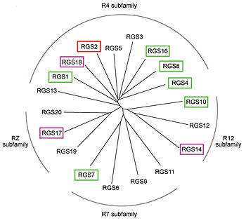
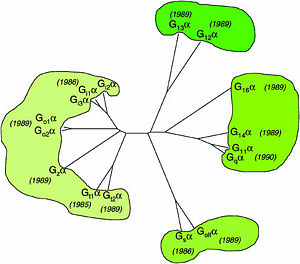
Like many signaling proteins, RGS proteins comprise a large and diverse family. In human genome, Thirty-seven RGS proteins are encoded by gene loci; this collection of related proteins has been divided into 10 different subfamilies based on the relatedness of their RGS domain sequence and their multiple domain architectures. About 20 ‘canonical’ RGS proteins can in theory downregulate any of the 16 activated Gα subunits, although in practice they interact only with members of the G<sub>i</sub> and G<sub>q</sub> families. In these proteins, the ~120-residue RGS homology domain functions as a GTPase-activating protein (GAP) for GTP-bound Gα subunits. | Like many signaling proteins, RGS proteins comprise a large and diverse family. In human genome, Thirty-seven RGS proteins are encoded by gene loci; this collection of related proteins has been divided into 10 different subfamilies based on the relatedness of their RGS domain sequence and their multiple domain architectures. About 20 ‘canonical’ RGS proteins can in theory downregulate any of the 16 activated Gα subunits, although in practice they interact only with members of the G<sub>i</sub> and G<sub>q</sub> families. In these proteins, the ~120-residue RGS homology domain functions as a GTPase-activating protein (GAP) for GTP-bound Gα subunits. | ||
| Line 30: | Line 30: | ||
== Structural highlights == | == Structural highlights == | ||
| - | 1AGR is complex of RGS4 and Gα<sub>i1</sub> defined by X-ray crystallographic analysis. | + | 1AGR is complex of RGS4 and Gα<sub>i1</sub> proteins defined by X-ray crystallographic analysis. |
| - | <scene name='70/701447/Rgs4_monomer/4'> | + | <scene name='70/701447/Rgs4_monomer/4'> The RGS4 domain </scene> corresponds to an array of nine α-helices that fold into two small subdomains, both subdomains are required for his GAP activity. α1, α2, α3, α4, α5 and α6 helices colred blue, aqua, yellow, coral, magenta and dark green respectivly. the helices α7, α8 and α9 colored red. |
Gα<sub>i1</sub> subunits adopt a conserved fold composed of <scene name='70/701447/All-helical-domain/6'>α helical domain</scene> , a helical domain of six α helices shown as blue cartoon and a GTPase domain shown in gray cartoons. The GTPase domain hydrolyzes GTP and provides most of Gα's binding surfaces for Gβγ, receptors, effectors and RGS proteins. <scene name='70/701447/Gi-rgs4/19'>The GTPase domain</scene> contains three flexible regions designated switch-I presented as blue sticks, switch-II presented as magenta sticks and switch-III presented as green sticks that change conformation in response to GTP binding and hydrolysis, GDP–Mg<sup>+2</sup>, bound in the active site of Gα<sub>i1</sub> is shown as a ball-and-stick model. The three switch regions of Gα<sub>i1</sub>: residues 176–184, 201–215, and 233–241, respectively . <ref>PMID: 9108480</ref> | Gα<sub>i1</sub> subunits adopt a conserved fold composed of <scene name='70/701447/All-helical-domain/6'>α helical domain</scene> , a helical domain of six α helices shown as blue cartoon and a GTPase domain shown in gray cartoons. The GTPase domain hydrolyzes GTP and provides most of Gα's binding surfaces for Gβγ, receptors, effectors and RGS proteins. <scene name='70/701447/Gi-rgs4/19'>The GTPase domain</scene> contains three flexible regions designated switch-I presented as blue sticks, switch-II presented as magenta sticks and switch-III presented as green sticks that change conformation in response to GTP binding and hydrolysis, GDP–Mg<sup>+2</sup>, bound in the active site of Gα<sub>i1</sub> is shown as a ball-and-stick model. The three switch regions of Gα<sub>i1</sub>: residues 176–184, 201–215, and 233–241, respectively . <ref>PMID: 9108480</ref> | ||
Revision as of 21:13, 18 May 2015
Regulator of G protein signaling (RGS) interactions with G proteins – RGS4-Gαi as a model structure.
| |||||||||||


References
- ↑ Kosloff M, Travis AM, Bosch DE, Siderovski DP, Arshavsky VY. Integrating energy calculations with functional assays to decipher the specificity of G protein-RGS protein interactions. Nat Struct Mol Biol. 2011 Jun 19;18(7):846-53. doi: 10.1038/nsmb.2068. PMID:21685921 doi:http://dx.doi.org/10.1038/nsmb.2068
- ↑ Milligan G, Kostenis E. Heterotrimeric G-proteins: a short history. Br J Pharmacol. 2006 Jan;147 Suppl 1:S46-55. PMID:16402120 doi:http://dx.doi.org/10.1038/sj.bjp.0706405
- ↑ Tesmer JJ, Berman DM, Gilman AG, Sprang SR. Structure of RGS4 bound to AlF4--activated G(i alpha1): stabilization of the transition state for GTP hydrolysis. Cell. 1997 Apr 18;89(2):251-61. PMID:9108480
Proteopedia Page Contributors and Editors (what is this?)
Ali Asli, Denise Salem, Michal Harel, Joel L. Sussman, Jaime Prilusky
