We apologize for Proteopedia being slow to respond. For the past two years, a new implementation of Proteopedia has been being built. Soon, it will replace this 18-year old system. All existing content will be moved to the new system at a date that will be announced here.
Talk:SCF-c-Kit
From Proteopedia
(Difference between revisions)
| Line 5: | Line 5: | ||
KIT was initially discovered as an oncogene in a feline retrovirus. | KIT was initially discovered as an oncogene in a feline retrovirus. | ||
SCF and KIT are required for a normal development of hematopoietic cells, melanocytes and others. In humans, loss-of-function mutations in KIT cause thepiebald trait. A variety of gain-of-function mutations in KIT were found in different types of human cancers such as gastro-intestinal-stromal tumors, acute myeloid leukemia, and mast cell leukemia. | SCF and KIT are required for a normal development of hematopoietic cells, melanocytes and others. In humans, loss-of-function mutations in KIT cause thepiebald trait. A variety of gain-of-function mutations in KIT were found in different types of human cancers such as gastro-intestinal-stromal tumors, acute myeloid leukemia, and mast cell leukemia. | ||
| + | |||
You may include any references to papers as in: the use of JSmol in Proteopedia <ref>DOI 10.1002/ijch.201300024</ref> or to the article describing Jmol <ref>PMID:21638687</ref> to the rescue. | You may include any references to papers as in: the use of JSmol in Proteopedia <ref>DOI 10.1002/ijch.201300024</ref> or to the article describing Jmol <ref>PMID:21638687</ref> to the rescue. | ||
| Line 26: | Line 27: | ||
== mutations == | == mutations == | ||
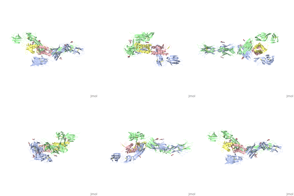
A point mutation in either Arg381 or Glu386 within D4 can strongly compromise SCF-induced tyrosine autophosphorylation. The homotypic interactions between membrane-proximal regions of KIT are mediated primarily by the D4-D4 interface, while the D5-D5 interface plays a cooperative secondary role, i.e., the D5-D5 faciliates the exact positioning of two KIT ectodomains at the cellsurface interface. SCF-KIT binding occurs in at least two steps: first, the electrostatic attraction between SCF and D1-D2-D3 will align SCF along the opposing ligand-binding region on KIT. A faster association rate might occur as a result of an electrostatic attraction of SCF due to a steering effect. Interestingly, the main interactions that maintain the D4-D4 interface, i.e. double salt bridges between Arg381 and Glu386 in a neighboring KIT molecule are also mediated by electrostatic interactions. Cell-cell interactions, the ectodomain of KIT has evolved since the hallmarks of KIT structure, ligand binding, and receptor dimerization are conserved in other receptors, the mechanism described above for KIT activation may be a general mechanism for activation of many receptors. | A point mutation in either Arg381 or Glu386 within D4 can strongly compromise SCF-induced tyrosine autophosphorylation. The homotypic interactions between membrane-proximal regions of KIT are mediated primarily by the D4-D4 interface, while the D5-D5 interface plays a cooperative secondary role, i.e., the D5-D5 faciliates the exact positioning of two KIT ectodomains at the cellsurface interface. SCF-KIT binding occurs in at least two steps: first, the electrostatic attraction between SCF and D1-D2-D3 will align SCF along the opposing ligand-binding region on KIT. A faster association rate might occur as a result of an electrostatic attraction of SCF due to a steering effect. Interestingly, the main interactions that maintain the D4-D4 interface, i.e. double salt bridges between Arg381 and Glu386 in a neighboring KIT molecule are also mediated by electrostatic interactions. Cell-cell interactions, the ectodomain of KIT has evolved since the hallmarks of KIT structure, ligand binding, and receptor dimerization are conserved in other receptors, the mechanism described above for KIT activation may be a general mechanism for activation of many receptors. | ||
| + | |||
| + | <Structure load='2E9W' size='350' frame='true' align='right' caption='Insert caption here' scene='Insert optional scene name here' /> | ||
This is a sample scene created with SAT to <scene name="/12/3456/Sample/1">color</scene> by Group, and another to make <scene name="/12/3456/Sample/2">a transparent representation</scene> of the protein. You can make your own scenes on SAT starting from scratch or loading and editing one of these sample scenes. | This is a sample scene created with SAT to <scene name="/12/3456/Sample/1">color</scene> by Group, and another to make <scene name="/12/3456/Sample/2">a transparent representation</scene> of the protein. You can make your own scenes on SAT starting from scratch or loading and editing one of these sample scenes. | ||
Revision as of 11:31, 24 May 2015
Introduction)
| |||||||||||

