This old version of Proteopedia is provided for student assignments while the new version is undergoing repairs. Content and edits done in this old version of Proteopedia after March 1, 2026 will eventually be lost when it is retired in about June of 2026.
Apply for new accounts at the new Proteopedia. Your logins will work in both the old and new versions.
Beta2 adrenergic receptor-Gs protein complex
From Proteopedia
(Difference between revisions)
| Line 8: | Line 8: | ||
== Complex structure == | == Complex structure == | ||
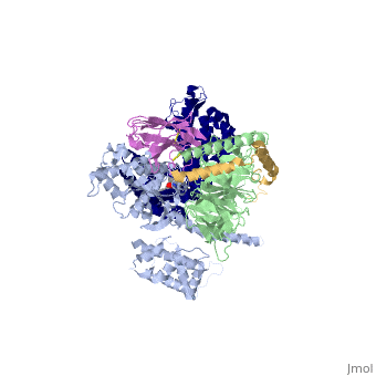
| - | The overall structure shows the | + | The overall structure shows the β2AR (dark blue) bound to an agonist (in spheres) along with a T4 lysozyme fused to its amino terminus in order to facilitate crystallization. The receptor interacts with Gαs (light blue). Gαs together with Gβ (light green) and Gγ (gold) constitute the heterotrimeric G protein Gs. A Gs-binding nanobody which also facilitates crystallization (pink) binds the G protein between the a and b subunits. |
== G-Protein-GPCR Intercations == | == G-Protein-GPCR Intercations == | ||
| - | The | + | The α5-helix of Gαs docks into a cavity formed on the intracellular side of the receptor by the opening of transmembrane helices 5 and 6. Within the transmembrane core, the interactions are primarily non-polar - an exception involves <scene name='70/701430/Receptor_g_protein_interaction/4'>packing of Tyr 391 of the α5-helix against Arg 131 of the conserved DRY sequence in TM3. Arg 131 also packs against Tyr 326 of the conserved NPxxY sequence in TM7</scene>. As the α5-helix exits the receptor it forms a network of polar interactions with TM5 and TM3. Receptor residues <scene name='70/701430/Receptor_gprotein_interaction2/1'>Thr 68 and Asp 130 interact with the ICL2 helix of the β2AR via Tyr 141, positioning the helix so that Phe 139 of the receptor docks into a hydrophobic pocket on the G protein surface</scene>, thereby structurally linking receptor–G protein interactions with the highly conserved DRY motif of the β2AR. |
== G-Protein Cycle == | == G-Protein Cycle == | ||
| - | [[Image:ImgSmall1.jpg|500px|G protein cycle for the | + | [[Image:ImgSmall1.jpg|500px|G protein cycle for the β2AR–Gs complex. Reprinted by permission from Macmillan Publishers Ltd on behalf of Cancer Research UK: Nature 477, 549–555, copyright 2011]] |
The figure shows the G Protein cycle <ref>doi:10.1038/nature10361</ref> - an extracellular agonist binding to the β2AR leads <scene name='70/701430/Receptor_morphing_animation/2'>to conformational rearrangements of the cytoplasmic ends of transmembrane segments</scene> that enable the Gs heterotrimer to bind the receptor. GDP is released from the α subunit upon formation of β2AR–Gs complex. The GTP binds to the nucleotide-free α subunit resulting in dissociation of the α and βγ subunits from the receptor. The subunits regulate their respective effector proteins adenylyl cyclase (AC) and Ca2+ channels. The Gs heterotrimer reassembles from α and βγ subunits following hydrolysis of GTP to GDP in the α subunit. | The figure shows the G Protein cycle <ref>doi:10.1038/nature10361</ref> - an extracellular agonist binding to the β2AR leads <scene name='70/701430/Receptor_morphing_animation/2'>to conformational rearrangements of the cytoplasmic ends of transmembrane segments</scene> that enable the Gs heterotrimer to bind the receptor. GDP is released from the α subunit upon formation of β2AR–Gs complex. The GTP binds to the nucleotide-free α subunit resulting in dissociation of the α and βγ subunits from the receptor. The subunits regulate their respective effector proteins adenylyl cyclase (AC) and Ca2+ channels. The Gs heterotrimer reassembles from α and βγ subunits following hydrolysis of GTP to GDP in the α subunit. | ||
| Line 20: | Line 20: | ||
== G-Protein variability == | == G-Protein variability == | ||
| - | The Gαs subunit consists of two domains, the Ras domain (αRas) and the α-helical domain (αAH). Both are involved in nucleotide binding. A big discovery made thanks to this structure is that the | + | The Gαs subunit consists of two domains, the Ras domain (αRas) and the α-helical domain (αAH). Both are involved in nucleotide binding. A big discovery made thanks to this structure is that the Gα subunit has large variability between its GTP bound (active) and nucleotide free states, where <scene name='70/701430/Gamorph/2'>the αAH domain has a variable position relative to the αRas domain</scene> |
==See Also== | ==See Also== | ||
Revision as of 19:16, 3 June 2015
Beta2 adrenergic receptor-Gs protein complex
| |||||||||||
References
- ↑ Rasmussen SG, DeVree BT, Zou Y, Kruse AC, Chung KY, Kobilka TS, Thian FS, Chae PS, Pardon E, Calinski D, Mathiesen JM, Shah ST, Lyons JA, Caffrey M, Gellman SH, Steyaert J, Skiniotis G, Weis WI, Sunahara RK, Kobilka BK. Crystal structure of the beta2 adrenergic receptor-Gs protein complex. Nature. 2011 Jul 19;477(7366):549-55. doi: 10.1038/nature10361. PMID:21772288 doi:10.1038/nature10361
Proteopedia Page Contributors and Editors (what is this?)
Dan Elran, Michal Harel, Alexander Berchansky, Joel L. Sussman
