Sandbox Reserved 1051
From Proteopedia
(Difference between revisions)
| Line 19: | Line 19: | ||
==Structure== | ==Structure== | ||
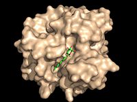
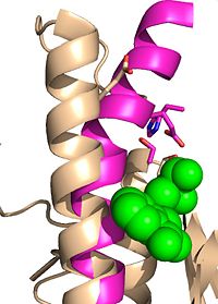
[[Image:Substrate Binding 2D Surface.jpg|200 px|left|thumb|'''Figure 2:''' Octylthioglucoside (green), a substrate analog, shown in the binding pocket of Ag85C (tan, from [[1va5]])]] | [[Image:Substrate Binding 2D Surface.jpg|200 px|left|thumb|'''Figure 2:''' Octylthioglucoside (green), a substrate analog, shown in the binding pocket of Ag85C (tan, from [[1va5]])]] | ||
| + | [[Image:Octylthioglucoside.jpg|200 px|left|thumb|'''Figure 3:''' Octylthioglucoside]] | ||
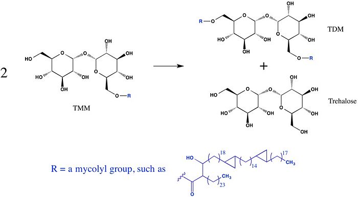
Wild type Ag85C, in the absence of any specific ligands, was originally crystallized as a <scene name='69/694219/Dimer/1'>homodimer</scene>.<ref name="Ronning2000">PMID: 10655617</ref> The <scene name='69/694220/Secondary_structures/2'>secondary structure</scene> (shown in the monomeric form) is 38% α-helical (pink) and 16% β-sheet (yellow). The confrontation of a central β-sheet bordered by α–helices comprises an [http://en.wikipedia.org/wiki/Alpha/beta_hydrolase_fold α/β hydrolase fold] in Ag85C, a supersecondary structure that is highly conserved across serine hydrolases. The active site of Ag85C is composed of two adjoined binding pockets; there is carbohydrate binding pocket for the trehalose substrate, and there is a fatty acid binding pocket for the mycolic acid. As a result, trehalose monomycolate can effectively bind to the Ag85C active site. This binding pocket is shown in Figure 2, in which Ag85C is shown with a bound substrate mimic, octylthioglucoside<ref name="Ronning2004">PMID: 15192106</ref> (Figure 3). | Wild type Ag85C, in the absence of any specific ligands, was originally crystallized as a <scene name='69/694219/Dimer/1'>homodimer</scene>.<ref name="Ronning2000">PMID: 10655617</ref> The <scene name='69/694220/Secondary_structures/2'>secondary structure</scene> (shown in the monomeric form) is 38% α-helical (pink) and 16% β-sheet (yellow). The confrontation of a central β-sheet bordered by α–helices comprises an [http://en.wikipedia.org/wiki/Alpha/beta_hydrolase_fold α/β hydrolase fold] in Ag85C, a supersecondary structure that is highly conserved across serine hydrolases. The active site of Ag85C is composed of two adjoined binding pockets; there is carbohydrate binding pocket for the trehalose substrate, and there is a fatty acid binding pocket for the mycolic acid. As a result, trehalose monomycolate can effectively bind to the Ag85C active site. This binding pocket is shown in Figure 2, in which Ag85C is shown with a bound substrate mimic, octylthioglucoside<ref name="Ronning2004">PMID: 15192106</ref> (Figure 3). | ||
| - | INSERT CHEMDRAW PICTURE COMPARING THE OCTYLTHIOGLYCOSIDE STRUCTURE TO TMM | ||
===Catalytic Triad=== | ===Catalytic Triad=== | ||
Revision as of 14:28, 10 June 2015
| This Sandbox is Reserved from 02/09/2015, through 05/31/2016 for use in the course "CH462: Biochemistry 2" taught by Geoffrey C. Hoops at the Butler University. This reservation includes Sandbox Reserved 1051 through Sandbox Reserved 1080. |
To get started:
More help: Help:Editing |
Trehalose-O-mycolyltransferase Ag85C
Introduction
Antigen 85C is one of three homologous protein components of the Ag85 complex in the cell wall of M. tuberculosis. This serine esterase enzyme catalyzes the transfer of mycolyl groups, characteristic components of the cell wall of mycobacteria. Several three dimensional structures of Ag85C have been solved, including the wild type enzyme as well as active site variants due to site-directed mutagenesis and covalent modification.
| |||||||||||
References
- ↑ Jackson M, Raynaud C, Laneelle MA, Guilhot C, Laurent-Winter C, Ensergueix D, Gicquel B, Daffe M. Inactivation of the antigen 85C gene profoundly affects the mycolate content and alters the permeability of the Mycobacterium tuberculosis cell envelope. Mol Microbiol. 1999 Mar;31(5):1573-87. PMID:10200974
- ↑ Ronning DR, Klabunde T, Besra GS, Vissa VD, Belisle JT, Sacchettini JC. Crystal structure of the secreted form of antigen 85C reveals potential targets for mycobacterial drugs and vaccines. Nat Struct Biol. 2000 Feb;7(2):141-6. PMID:10655617 doi:10.1038/72413
- ↑ Ronning DR, Vissa V, Besra GS, Belisle JT, Sacchettini JC. Mycobacterium tuberculosis antigen 85A and 85C structures confirm binding orientation and conserved substrate specificity. J Biol Chem. 2004 Aug 27;279(35):36771-7. Epub 2004 Jun 10. PMID:15192106 doi:http://dx.doi.org/10.1074/jbc.M400811200
- ↑ Favrot L, Lajiness DH, Ronning DR. Inactivation of the Mycobacterium tuberculosis Antigen 85 complex by covalent, allosteric inhibitors. J Biol Chem. 2014 Jul 14. pii: jbc.M114.582445. PMID:25028518 doi:http://dx.doi.org/10.1074/jbc.M114.582445
