We apologize for Proteopedia being slow to respond. For the past two years, a new implementation of Proteopedia has been being built. Soon, it will replace this 18-year old system. All existing content will be moved to the new system at a date that will be announced here.
PLC beta 3 Gq
From Proteopedia
(Difference between revisions)
| Line 21: | Line 21: | ||
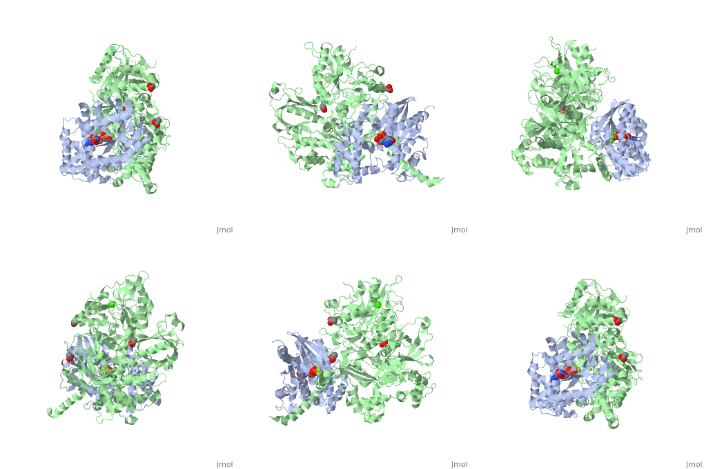
<scene name='70/701452/Fig7/1'>An extended loop</scene> (residues 254-268) between EF hands 3 and 4 of PLC- β3 interacts with the GTP-binding region of Gαq. Asn260 of the EF3/4 loop promotes GTP hydrolysis by interaction with the side chain of Gln209 of Gαq, which rearranges during GTP hydrolysis to stabilize the transition state mimicked by GDP•AlF4–•H20. Asn260 also interacts with Glu212 to stabilize switch 1 for GTP hydrolysis. | <scene name='70/701452/Fig7/1'>An extended loop</scene> (residues 254-268) between EF hands 3 and 4 of PLC- β3 interacts with the GTP-binding region of Gαq. Asn260 of the EF3/4 loop promotes GTP hydrolysis by interaction with the side chain of Gln209 of Gαq, which rearranges during GTP hydrolysis to stabilize the transition state mimicked by GDP•AlF4–•H20. Asn260 also interacts with Glu212 to stabilize switch 1 for GTP hydrolysis. | ||
| - | Other effectors are known to engage residues (pink) in the <scene name='70/701452/Fig3/3'>effector-binding site</scene> within Gα subunits. There are a large family of regulator of G protein signaling (RGS) proteins that independently accelerates the GTP hydrolysis in the <scene name='70/701452/Fig4/3'>GTPase domain</scene> composed out of several residues (brown). In comparison to PLC-β3 interacts with residues (blue) on Gαq that <scene name='70/701452/Fig5/4'>overlaps almost completely</scene> with portions of Gα subunits needed for engagement of RGS proteins and the effector-binding region. | + | Other effectors are known to engage residues (pink) in the <scene name='70/701452/Fig3/3'>effector-binding site</scene> <ref>PMID:20966218</ref> within Gα subunits. There are a large family of regulator of G protein signaling (RGS) proteins that independently accelerates the GTP hydrolysis in the <scene name='70/701452/Fig4/3'>GTPase domain</scene> composed out of several residues (brown). In comparison to PLC-β3 interacts with residues (blue) on Gαq that <scene name='70/701452/Fig5/4'>overlaps almost completely</scene> with portions of Gα subunits needed for engagement of RGS proteins and the effector-binding region. |
'''Critical residues in the interface:''' | '''Critical residues in the interface:''' | ||
Revision as of 09:18, 18 July 2015
Unique bidirectional interactions of Phospholipase C beta 3 with G alpha Q
| |||||||||||
References
- ↑ Waldo GL, Ricks TK, Hicks SN, Cheever ML, Kawano T, Tsuboi K, Wang X, Montell C, Kozasa T, Sondek J, Harden TK. Kinetic Scaffolding Mediated by a Phospholipase C-{beta} and Gq Signaling Complex. Science. 2010 Nov 12;330(6006):974-80. Epub 2010 Oct 21. PMID:20966218 doi:10.1126/science.1193438
- ↑ Lyon AM, Tesmer JJ. Structural insights into phospholipase C-beta function. Mol Pharmacol. 2013 Oct;84(4):488-500. doi: 10.1124/mol.113.087403. Epub 2013 Jul, 23. PMID:23880553 doi:http://dx.doi.org/10.1124/mol.113.087403
- ↑ Waldo GL, Ricks TK, Hicks SN, Cheever ML, Kawano T, Tsuboi K, Wang X, Montell C, Kozasa T, Sondek J, Harden TK. Kinetic Scaffolding Mediated by a Phospholipase C-{beta} and Gq Signaling Complex. Science. 2010 Nov 12;330(6006):974-80. Epub 2010 Oct 21. PMID:20966218 doi:10.1126/science.1193438

