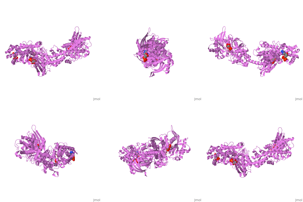
1cza
From Proteopedia
| Line 4: | Line 4: | ||
|PDB= 1cza |SIZE=350|CAPTION= <scene name='initialview01'>1cza</scene>, resolution 1.9Å | |PDB= 1cza |SIZE=350|CAPTION= <scene name='initialview01'>1cza</scene>, resolution 1.9Å | ||
|SITE= | |SITE= | ||
| - | |LIGAND= <scene name='pdbligand=GLC:GLUCOSE'>GLC</scene>, <scene name='pdbligand=G6P:ALPHA-D-GLUCOSE-6-PHOSPHATE'>G6P</scene> and <scene name='pdbligand=ADP:ADENOSINE-5 | + | |LIGAND= <scene name='pdbligand=GLC:GLUCOSE'>GLC</scene>, <scene name='pdbligand=G6P:ALPHA-D-GLUCOSE-6-PHOSPHATE'>G6P</scene> and <scene name='pdbligand=ADP:ADENOSINE-5'-DIPHOSPHATE'>ADP</scene> |
|ACTIVITY= [http://en.wikipedia.org/wiki/Hexokinase Hexokinase], with EC number [http://www.brenda-enzymes.info/php/result_flat.php4?ecno=2.7.1.1 2.7.1.1] | |ACTIVITY= [http://en.wikipedia.org/wiki/Hexokinase Hexokinase], with EC number [http://www.brenda-enzymes.info/php/result_flat.php4?ecno=2.7.1.1 2.7.1.1] | ||
|GENE= | |GENE= | ||
| Line 38: | Line 38: | ||
[[Category: structurally homologous domain]] | [[Category: structurally homologous domain]] | ||
| - | ''Page seeded by [http://oca.weizmann.ac.il/oca OCA ] on | + | ''Page seeded by [http://oca.weizmann.ac.il/oca OCA ] on Sun Mar 23 11:26:36 2008'' |
Revision as of 09:26, 23 March 2008
| |||||||
| , resolution 1.9Å | |||||||
|---|---|---|---|---|---|---|---|
| Ligands: | , and | ||||||
| Activity: | Hexokinase, with EC number 2.7.1.1 | ||||||
| Coordinates: | save as pdb, mmCIF, xml | ||||||
MUTANT MONOMER OF RECOMBINANT HUMAN HEXOKINASE TYPE I COMPLEXED WITH GLUCOSE, GLUCOSE-6-PHOSPHATE, AND ADP
Contents |
Overview
Hexokinase I, the pacemaker of glycolysis in brain tissue, is composed of two structurally similar halves connected by an alpha-helix. The enzyme dimerizes at elevated protein concentrations in solution and in crystal structures; however, almost all published data reflect the properties of a hexokinase I monomer in solution. Crystal structures of mutant forms of recombinant human hexokinase I, presented here, reveal the enzyme monomer for the first time. The mutant hexokinases bind both glucose 6-phosphate and glucose with high affinity to their N and C-terminal halves, and ADP, also with high affinity, to a site near the N terminus of the polypeptide chain. Exposure of the monomer crystals to ADP in the complete absence of glucose 6-phosphate reveals a second binding site for adenine nucleotides at the putative active site (C-half), with conformational changes extending 15 A to the contact interface between the N and C-halves. The structures reveal distinct conformational states for the C-half and a rigid-body rotation of the N-half, as possible elements of a structure-based mechanism for allosteric regulation of catalysis.
Disease
Known disease associated with this structure: Hemolytic anemia due to hexokinase deficiency OMIM:[142600]
About this Structure
1CZA is a Single protein structure of sequence from Homo sapiens. Full crystallographic information is available from OCA.
Reference
Crystal structures of mutant monomeric hexokinase I reveal multiple ADP binding sites and conformational changes relevant to allosteric regulation., Aleshin AE, Kirby C, Liu X, Bourenkov GP, Bartunik HD, Fromm HJ, Honzatko RB, J Mol Biol. 2000 Mar 3;296(4):1001-15. PMID:10686099
Page seeded by OCA on Sun Mar 23 11:26:36 2008

