This old version of Proteopedia is provided for student assignments while the new version is undergoing repairs. Content and edits done in this old version of Proteopedia after March 1, 2026 will eventually be lost when it is retired in about June of 2026.
Apply for new accounts at the new Proteopedia. Your logins will work in both the old and new versions.
Sanbox glut3
From Proteopedia
(Difference between revisions)
| Line 5: | Line 5: | ||
== Function == | == Function == | ||
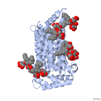
| - | <scene name='71/716527/5c65/1'>GLUT3</scene> is one of fourteen facilitative sugar transporters which use the glucose diffusion gradient to move across various plasma membranes to display various specificities, kinetics and tissue expression profiles<ref> | + | <scene name='71/716527/5c65/1'>GLUT3</scene> is one of fourteen facilitative sugar transporters which use the glucose diffusion gradient to move across various plasma membranes to display various specificities, kinetics and tissue expression profiles<ref> Structure of, and functional insight into the GLUT family of membrane transporters. Cell Health and Cytoskeleton, 7, 167-183. doi:10.2147/CHC.S60484</ref> (Long et al. 2015). Glucose transporters are about 500 amino acids in length and are a part of a growing superfamily of integral membrane glycoproteins that have 12 transmembrane (TM) helices. The transmembrane regions presumably create channels through which glucose can move (Kipmen-Korgun et al. 2009). GLUT3 is categorized as a Class I transporter due to its protein sequence and structural similarity to other glucose transporters grouped in Class I (Long et al. 2015). GLUT3 displays the highest affinity for glucose of all of the Class I glucose transporters and has a transport capacity five times greater than that of GLUT1 and GLUT4 (Simpson et al., 2008). In humans, GLUT3 is found predominantly in brain tissue, highly and specifically expressed by neurons, and has some expression in peripheral tissues (Maher et al., 1994; Simpson et al. 2008). It is commonly known as the “neuronal glucose transporter” because of it is most commonly found in the brain, specifically seen mostly in the axons and dendrites of neurons (Simpson et al. 2008). GLUT3 has a more restricted expression pathway, which represents specialized functions for the protein (Xu et al., 2015). GLUT3 has been found to play an important role in gestational development and maintaining the brain's structure. Defects in GLUT3 can cause fetal death as well as neurodegeneration, which can lead to diseases like Alzheimer’s (Liu et al. and Zu et al. 2008). |
==Structure== | ==Structure== | ||
Revision as of 03:16, 17 November 2015
Facilitated Glucose Transporter 3, Solute Carrier Family 2 (GLUT3/ SLC2A3) in Homo Sapiens
| |||||||||||
References
- ↑ Hanson, R. M., Prilusky, J., Renjian, Z., Nakane, T. and Sussman, J. L. (2013), JSmol and the Next-Generation Web-Based Representation of 3D Molecular Structure as Applied to Proteopedia. Isr. J. Chem., 53:207-216. doi:http://dx.doi.org/10.1002/ijch.201300024
- ↑ Herraez A. Biomolecules in the computer: Jmol to the rescue. Biochem Mol Biol Educ. 2006 Jul;34(4):255-61. doi: 10.1002/bmb.2006.494034042644. PMID:21638687 doi:10.1002/bmb.2006.494034042644
- ↑ Structure of, and functional insight into the GLUT family of membrane transporters. Cell Health and Cytoskeleton, 7, 167-183. doi:10.2147/CHC.S60484
