This old version of Proteopedia is provided for student assignments while the new version is undergoing repairs. Content and edits done in this old version of Proteopedia after March 1, 2026 will eventually be lost when it is retired in about June of 2026.
Apply for new accounts at the new Proteopedia. Your logins will work in both the old and new versions.
Sanbox glut3
From Proteopedia
(Difference between revisions)
| Line 12: | Line 12: | ||
== Disease in Humans == | == Disease in Humans == | ||
===Type 2 Diabetes=== | ===Type 2 Diabetes=== | ||
| - | Higher glucose concentration, as seen in diabetics, influences GLUT expression in leukocytes. Patients with type 2 diabetes have decreased GLUT3 in granulocytes, lymphocytes, and monocytes. In addition, the level of transcripts that encode GLUT3 are reduced in diabetic patients. Decreased expression of GLUT3 and other GLUT isoforms could possibly impair immune function and increase susceptibility to infection in type 2 diabetes | + | Higher glucose concentration, as seen in diabetics, influences GLUT expression in leukocytes. Patients with type 2 diabetes have decreased GLUT3 in granulocytes, lymphocytes, and monocytes. In addition, the level of transcripts that encode GLUT3 are reduced in diabetic patients. Decreased expression of GLUT3 and other GLUT isoforms could possibly impair immune function and increase susceptibility to infection in type 2 diabetes<ref name="four"/>. |
===Alzheimer's disease=== | ===Alzheimer's disease=== | ||
| - | Alzheimer’s disease shows levels of impaired glucose uptake and metabolism, which leads to the downgrade of many other factors in the brain. GLUT3 is responsible for transporting glucose from extracellular space to neuronal tissue, specifically dendrites and axons. Decreased levels of GLUT3 in Alzheimer brain shows a positive correlation to decreased levels of N-acetylglucosamine. The impaired presence of GLUT3 leads to hyperphosphorylation of the Tau protein, which normally stabilizes neuronal microtubules. Lastly there is a reduction in the transcription for factor hypoxia-inducible factor 1, which plays a role in glucose metabolism in the brain. The comparison between a normal healthy brain and an Alzheimer brain relieved that there was a 25-30% decrease in GLUT3 levels in the Alzheimer brain | + | Alzheimer’s disease shows levels of impaired glucose uptake and metabolism, which leads to the downgrade of many other factors in the brain. GLUT3 is responsible for transporting glucose from extracellular space to neuronal tissue, specifically dendrites and axons. Decreased levels of GLUT3 in Alzheimer brain shows a positive correlation to decreased levels of N-acetylglucosamine. The impaired presence of GLUT3 leads to hyperphosphorylation of the Tau protein, which normally stabilizes neuronal microtubules. Lastly there is a reduction in the transcription for factor hypoxia-inducible factor 1, which plays a role in glucose metabolism in the brain. The comparison between a normal healthy brain and an Alzheimer brain relieved that there was a 25-30% decrease in GLUT3 levels in the Alzheimer brain<ref name="eight"/>. |
===Huntington’s Disease=== | ===Huntington’s Disease=== | ||
| - | Huntington’s disease leads to decreased expression of GLUT3 in the plasma membrane. Increasing the expression of GLUT3 in a Huntington’s disease brain can delay the onset of the disease | + | Huntington’s disease leads to decreased expression of GLUT3 in the plasma membrane. Increasing the expression of GLUT3 in a Huntington’s disease brain can delay the onset of the disease<ref name="ten">Vittori, A., Breda, C., Repici, M., Orth, M., Roos, R. A. C., Outeiro, T. F., . . . the REGISTRY investigators of the European Huntington's Disease Network. (2014). Copy-number variation of the neuronal glucose transporter gene SLC2A3 and age of onset in huntington's disease. Human Molecular Genetics, 23(12), 3129-3137. doi:10.1093/hmg/ddu022</ref>. Rab11 is a protein that is involved with the regulation of transporter trafficking. It helps in the regulation of glucose transporters particularly the GLUT3 transporter. Its regulation is impaired by Huntington’s disease, which leads to the decreased cell surface expression of GLUT3 in the brain. The exact mechanism of Huntington’s disease is still unknown to this day<ref name="eleven">McClory, H., Williams, D., & Sapp, E. (2014). Glucose transporter 3 is a rab11-dependent trafficking cargo and its transport to the cell surface is reduced in neurons of CAG140 Huntington’s disease mice. Acta Neuropathol Commun, 2, 1-9.</ref>. |
==Mechanism == | ==Mechanism == | ||
Revision as of 05:41, 17 November 2015
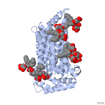
Facilitated Glucose Transporter 3, Solute Carrier Family 2 (GLUT3/ SLC2A3) in Homo Sapiens
| |||||||||||
References
- ↑ 1.0 1.1 1.2 1.3 Long, W., & Cheeseman, C. I. (2015). Structure of, and functional insight into the GLUT family of membrane transporters. Cell Health and Cytoskeleton, 7, 167-183. doi:10.2147/CHC.S60484
- ↑ 2.0 2.1 Kipmen-Korgun, D., Bilmen-Sarikcioglu, S., Altunbas, H., Demir, R., & Korgun, E. T. (2009). Type-2 diabetes down-regulates glucose transporter proteins and genes of the human blood leukocytes.Scandinavian Journal of Clinical and Laboratory Investigation, 69(3), 350-358. doi:10.1080/00365510802632163
- ↑ 3.0 3.1 Simpson,I. A., Dwyer, D., Malide, D., Moley, K. H., Travis, A., & Vannucci, S. J. (2008). The facilitative glucose transporter GLUT3: 20 years of distinction. American Journal of Physiology - Endocrinology and Metabolism, 295(2), E242-E253. doi:10.1152/ajpendo.90388.2008
- ↑ Maher, F., Vannucci, S. J., & Simpson, I. A. (1994). Glucose transporter proteins in brain. FASEB Journal, 8(13), 1003-1011.
- ↑ Xu, J., Lu, C., Wang, J., Zhang, R., Qian, X., & Zhu, H. (2015). Regulation of human trophoblast GLUT3 glucose transporter by mammalian target of rapamycin signaling. International Journal of Molecular Sciences, 16(6), 13815-13828. doi:10.3390/ijms160613815
- ↑ 6.0 6.1 Liu, Y., Liu, F., Iqbal, K., Grundke-Iqbal, I., & Gong, C. -. (2008). Decreased glucose transporters correlate to abnormal hyperphosphorylation of tau in alzheimer disease. FEBS Letters, 582(2), 359-364. doi:10.1016/j.febslet.2007.12.035
- ↑ 7.0 7.1 http://www.ebi.ac.uk/pdbe/entry/pdb/5c65/
- ↑ http://oca.weizmann.ac.il/oca-bin/ocaids?id=5c65
- ↑ 9.0 9.1 9.2 9.3 Deng, D., Sun, P., Yan, C., Ke, M., Jiang, X., Xiong, L., . . . Yan, N. (2015). Molecular basis of ligand recognition and transport by glucose transporters. Nature, 526(7573), 391-396. doi:10.1038/nature14655
- ↑ http://www.rcsb.org/pdb/explore.do?structureId=5C65
- ↑ http://www.ebi.ac.uk/pdbe/entry/pdb/5c65/bound/37X
- ↑ http://www.ebi.ac.uk/pdbe/entry/pdb/5c65/bound/Y01
- ↑ Vittori, A., Breda, C., Repici, M., Orth, M., Roos, R. A. C., Outeiro, T. F., . . . the REGISTRY investigators of the European Huntington's Disease Network. (2014). Copy-number variation of the neuronal glucose transporter gene SLC2A3 and age of onset in huntington's disease. Human Molecular Genetics, 23(12), 3129-3137. doi:10.1093/hmg/ddu022
- ↑ McClory, H., Williams, D., & Sapp, E. (2014). Glucose transporter 3 is a rab11-dependent trafficking cargo and its transport to the cell surface is reduced in neurons of CAG140 Huntington’s disease mice. Acta Neuropathol Commun, 2, 1-9.
- ↑ 15.0 15.1 Naftalin RJ, Holman GD. Transport of sugars in human red cells. In: Ellory JC, Lew V, editors. \ Membrane Transport in Red Cells. New York, NY, USA: Academic Press; 1977.
- ↑ 16.0 16.1 Carruthers, A., DeZutter, J., Ganguly, A., & Devaskar, S. U. (2009). Will the original glucose transporter isoform please stand up! American Journal of Physiology - Endocrinology and Metabolism, 297(4), E836-E848. doi:10.1152/ajpendo.00496.2009
- ↑ Jardetzky, O. (1966). Simple allosteric model for membrane pumps [27]. Nature, 211(5052), 969-970. doi:10.1038/211969a0
- ↑ Abramson J, Smirnova I, Kasho V, Verner G, Kaback HR, Iwata S. Structure and mechanism of the lactose permease of Escherichia coli. Science. 2003;301:610–615.
- ↑ Caulfield MJ, Munroe PB, O’Neill D, et al. SLC2A9 is a high-capacity urate transporter in humans. PLoS Med. 2008;5:1509–1523.
- ↑ Vollers, S. S., & Carruthers, A. (2012). Sequence determinants of GLUT1-mediated accelerated-exchange transport: Analysis by homology-scanning mutagenesis. Journal of Biological Chemistry, 287(51), 42533-42544.doi:10.1074/jbc.M112.369587
