This old version of Proteopedia is provided for student assignments while the new version is undergoing repairs. Content and edits done in this old version of Proteopedia after March 1, 2026 will eventually be lost when it is retired in about June of 2026.
Apply for new accounts at the new Proteopedia. Your logins will work in both the old and new versions.
Sanbox glut3
From Proteopedia
(Difference between revisions)
| Line 2: | Line 2: | ||
<StructureSection load='5c65' size='340' side='right' caption='GLUT3=''> | <StructureSection load='5c65' size='340' side='right' caption='GLUT3=''> | ||
== Function == | == Function == | ||
| - | GLUT3 is one of fourteen facilitative sugar transporters which use the glucose diffusion gradient to move across various plasma membranes to display various specificities, kinetics and tissue expression profiles <ref name="three">Long, W., & Cheeseman, C. I. (2015). Structure of, and functional insight into the GLUT family of membrane transporters. Cell Health and Cytoskeleton, 7, 167-183. doi:10.2147/CHC.S60484</ref>. Glucose transporters are approximately 500 amino acids in length and part of a growing superfamily of integral membrane glycoproteins that have 12 transmembrane (TM) helices. The transmembrane regions presumably create channels through which glucose can move<ref name="four">Kipmen-Korgun, D., Bilmen-Sarikcioglu, S., Altunbas, H., Demir, R., & Korgun, E. T. (2009). Type-2 diabetes down-regulates glucose transporter proteins and genes of the human blood leukocytes.Scandinavian Journal of Clinical and Laboratory Investigation, 69(3), 350-358. | + | GLUT3 is one of fourteen facilitative sugar transporters, which use the glucose diffusion gradient to move across various plasma membranes to display various specificities, kinetics and tissue expression profiles <ref name="three">Long, W., & Cheeseman, C. I. (2015). Structure of, and functional insight into the GLUT family of membrane transporters. Cell Health and Cytoskeleton, 7, 167-183. doi:10.2147/CHC.S60484</ref>. Glucose transporters are approximately 500 amino acids in length and part of a growing superfamily of integral membrane glycoproteins that have 12 transmembrane (TM) helices. The transmembrane regions presumably create channels through which glucose can move<ref name="four">Kipmen-Korgun, D., Bilmen-Sarikcioglu, S., Altunbas, H., Demir, R., & Korgun, E. T. (2009). Type-2 diabetes down-regulates glucose transporter proteins and genes of the human blood leukocytes.Scandinavian Journal of Clinical and Laboratory Investigation, 69(3), 350-358. |
doi:10.1080/00365510802632163</ref>. GLUT3 is categorized as a Class I transporter due to its protein sequence and structural similarity to other glucose transporters grouped in Class I<ref name="three"/>. GLUT3 displays the highest affinity for glucose of all of the Class I glucose transporters and has a transport capacity five times greater than that of GLUT1 and GLUT4<ref name="five"> Simpson,I. A., Dwyer, D., Malide, D., Moley, K. H., Travis, A., & Vannucci, S. J. (2008). The facilitative glucose transporter GLUT3: 20 years of distinction. American Journal of Physiology - Endocrinology and Metabolism, 295(2), E242-E253. doi:10.1152/ajpendo.90388.2008</ref>. In humans, GLUT3 is found predominantly in brain tissue, highly and specifically expressed by neurons, and has some expression in peripheral tissues. For this reason GLUT3 is commonly known as the “neuronal glucose transporter”<ref name="five"/><ref name="six">Maher, F., Vannucci, S. J., & Simpson, I. A. (1994). Glucose transporter proteins in brain. FASEB Journal, 8(13), 1003-1011.</ref>. GLUT3 has a more restricted expression pathway, which represents specialized functions for the protein<ref name="seven">Xu, J., Lu, C., Wang, J., Zhang, R., Qian, X., & Zhu, H. (2015). Regulation of human trophoblast GLUT3 glucose transporter by mammalian target of rapamycin signaling. International Journal of Molecular Sciences, 16(6), 13815-13828. doi:10.3390/ijms160613815</ref>. GLUT3 has been found to play an important role in gestational development and maintaining the brain's structure. Defects in GLUT3 can cause fetal death as well as neurodegeneration, which can lead to diseases like Alzheimer’s<ref name="eight">Liu, Y., Liu, F., Iqbal, K., Grundke-Iqbal, I., & Gong, C. -. (2008). Decreased glucose transporters correlate to abnormal hyperphosphorylation of tau in alzheimer disease. FEBS Letters, 582(2), 359-364. doi:10.1016/j.febslet.2007.12.035</ref>. | doi:10.1080/00365510802632163</ref>. GLUT3 is categorized as a Class I transporter due to its protein sequence and structural similarity to other glucose transporters grouped in Class I<ref name="three"/>. GLUT3 displays the highest affinity for glucose of all of the Class I glucose transporters and has a transport capacity five times greater than that of GLUT1 and GLUT4<ref name="five"> Simpson,I. A., Dwyer, D., Malide, D., Moley, K. H., Travis, A., & Vannucci, S. J. (2008). The facilitative glucose transporter GLUT3: 20 years of distinction. American Journal of Physiology - Endocrinology and Metabolism, 295(2), E242-E253. doi:10.1152/ajpendo.90388.2008</ref>. In humans, GLUT3 is found predominantly in brain tissue, highly and specifically expressed by neurons, and has some expression in peripheral tissues. For this reason GLUT3 is commonly known as the “neuronal glucose transporter”<ref name="five"/><ref name="six">Maher, F., Vannucci, S. J., & Simpson, I. A. (1994). Glucose transporter proteins in brain. FASEB Journal, 8(13), 1003-1011.</ref>. GLUT3 has a more restricted expression pathway, which represents specialized functions for the protein<ref name="seven">Xu, J., Lu, C., Wang, J., Zhang, R., Qian, X., & Zhu, H. (2015). Regulation of human trophoblast GLUT3 glucose transporter by mammalian target of rapamycin signaling. International Journal of Molecular Sciences, 16(6), 13815-13828. doi:10.3390/ijms160613815</ref>. GLUT3 has been found to play an important role in gestational development and maintaining the brain's structure. Defects in GLUT3 can cause fetal death as well as neurodegeneration, which can lead to diseases like Alzheimer’s<ref name="eight">Liu, Y., Liu, F., Iqbal, K., Grundke-Iqbal, I., & Gong, C. -. (2008). Decreased glucose transporters correlate to abnormal hyperphosphorylation of tau in alzheimer disease. FEBS Letters, 582(2), 359-364. doi:10.1016/j.febslet.2007.12.035</ref>. | ||
==Structure== | ==Structure== | ||
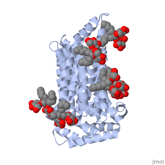
| - | GLUT3(<scene name='71/716527/5c65/1'>5c65</scene>) is a transport protein consisting of 481 amino acids and weighing 52,520 Daltons in its asymmetrical unit<ref name="nineteen">http://www.ebi.ac.uk/pdbe/entry/pdb/5c65/</ref>. This protein is an alpha-helical protein consisting of two chains, two different ligands and water<ref name="nineteen"/>. The structure was determined by X-Ray diffraction and was measured at a resolution of 2.65 Angstroms<ref name="twentytwo">http://oca.weizmann.ac.il/oca-bin/ocaids?id=5c65</ref>. GLUT3 consists of 12 transmembrane segments (TMs) folded “into the N-terminal and C-terminal domains, each comprising ‘3+3’ inverted repeats”<ref name="nine"/> These TMs consist of four 3 repeated sections. [http://www.nature.com/nature/journal/v526/n7573/fig_tab/nature14655_F1.html Here] is a figure by Deng, D., et al. showing these repeated transmembrane segments<ref name="nine">Deng, D., Sun, P., Yan, C., Ke, M., Jiang, X., Xiong, L., . . . Yan, N. (2015). Molecular basis of ligand recognition and transport by glucose transporters. Nature, 526(7573), 391-396. doi:10.1038/nature14655</ref>. The protein consists of two different ligands, Y01 | + | GLUT3(<scene name='71/716527/5c65/1'>5c65</scene>) is a transport protein consisting of 481 amino acids and weighing 52,520 Daltons in its asymmetrical unit<ref name="nineteen">http://www.ebi.ac.uk/pdbe/entry/pdb/5c65/</ref>. This protein is an alpha-helical protein consisting of two chains, two different ligands and water<ref name="nineteen"/>. The structure was determined by X-Ray diffraction and was measured at a resolution of 2.65 Angstroms<ref name="twentytwo">http://oca.weizmann.ac.il/oca-bin/ocaids?id=5c65</ref>. GLUT3 consists of 12 transmembrane segments (TMs) folded “into the N-terminal and C-terminal domains, each comprising ‘3+3’ inverted repeats”<ref name="nine"/> These TMs consist of four 3 repeated sections. [http://www.nature.com/nature/journal/v526/n7573/fig_tab/nature14655_F1.html Here] is a figure by Deng, D., et al. showing these repeated transmembrane segments<ref name="nine">Deng, D., Sun, P., Yan, C., Ke, M., Jiang, X., Xiong, L., . . . Yan, N. (2015). Molecular basis of ligand recognition and transport by glucose transporters. Nature, 526(7573), 391-396. doi:10.1038/nature14655</ref>. The protein consists of two different ligands, Y01 and 37X<ref name="eighteen">http://www.rcsb.org/pdb/explore.do?structureId=5C65</ref>. Octyl Glucose Neopentyl Glycol (<scene name='pdbligand=37X:OCTYL+GLUCOSE+NEOPENTYL+GLYCOL'>37X</scene>) has a chemical formula of C27H52O12 and a molecular weight of 569 Da. There are six 37X (501-506a) bound to chain A of 5c65. These ligands are kept in place by hydrogen bonds to arginine, proline, and serine and by van der Waals forces. Chain B has three 37X ligands attached to it (501-503b). These are attached through hydrogen bonds by arginine, proline, and serine as well as by van der Waals forces<ref name="twenty">http://www.ebi.ac.uk/pdbe/entry/pdb/5c65/bound/37X</ref>. To view 37X in 3D use [http://www.rcsb.org/pdb/explore/jmol.do?structureId=5C65&residueNr=37X JSmol]. Cholesterol hemisuccinate (<scene name='pdbligand=Y01:CHOLESTEROL+HEMISUCCINATE'>Y01</scene>) has a chemical formula of C31H50O4 and has a molecular weight of 487 Da. One Y01 is attached to chain a and another Y01 is attached to chain b<ref name="twentyone">http://www.ebi.ac.uk/pdbe/entry/pdb/5c65/bound/Y01</ref>. To view Y01 in 3D use [http://www.rcsb.org/pdb/explore/jmol.do?structureId=5C65&residueNr=Y01 JSmol]. GLUT3 was also identified and analyzed in a complex with alpha & beta d-glucose. This model was reported with a resolution of 1.5 Å and was in an open-occluded state<ref name="nine"/>. The alpha and beta d glucose were coordinated by amino acids N315, E378, Q159, W368, Q280, Q281, N286. These are located on TM8 and TM10a and TM10b<ref name="nine"/>. A figure of this glucose coordination by Deng, D., et al. is available [http://www.nature.com/nature/journal/v526/n7573/fig_tab/nature14655_F2.html here]. GLUT3 structure was also determined when bound to maltose in an outward-open and an outward-occluded conformation. This was measure to a resolution of 2.6 Å and 2.4 Å respectively. A figure of this maltose coordination by Deng, D., et al. is available [http://www.nature.com/nature/journal/v526/n7573/fig_tab/nature14655_F2.html here]. To get a better view of the structure of the protein use [http://oca.weizmann.ac.il/oca-docs/fgij/fg.htm?mol=5C65 FirstGlance]. |
This is 5c65 shown with <scene name="/12/3456/Sample/1">colored groups</scene>. This is 5c65 shown as a <scene name="/12/3456/Sample/2">transparent representation</scene> of the protein. | This is 5c65 shown with <scene name="/12/3456/Sample/1">colored groups</scene>. This is 5c65 shown as a <scene name="/12/3456/Sample/2">transparent representation</scene> of the protein. | ||
| Line 19: | Line 19: | ||
==Mechanism == | ==Mechanism == | ||
| - | Multiple mechanistic theories have been proposed for facilitated glucose transporters. The simple carrier model was the earliest theory proposed by Widdas and contains four steps. First, the empty carrier opens to the cis side of the membrane for glucose to bind<ref name="three"/>. Then the substrate binding carrier translocates to the trans side of the membrane where it then releases glucose on that side. Last the empty carrier switches to the cis side. Multiple mechanistic theories, including the simple carrier model were proposed but all attempted to explain two key components of GLUT transporters, the asymmetry of the transport affinities and the trans-acceleration that occurs in the presence of hexose on the trans side<ref name="twelve">Naftalin RJ, Holman GD. Transport of sugars in human red cells. In: Ellory JC, Lew V, editors. \ Membrane Transport in Red Cells. New York, NY, USA: Academic Press; 1977.</ref>. After considerable research, two popular models remain for class 1 glut transporters. The two-site/fixed site transporter theory explains the asymmetry by having both substrate binding sites simultaneously available<ref name="thirteen">Carruthers, A., DeZutter, J., Ganguly, A., & Devaskar, S. U. (2009). Will the original glucose transporter isoform please stand up! American Journal of Physiology - Endocrinology and Metabolism, 297(4), E836-E848. doi:10.1152/ajpendo.00496.2009 </ref>. After glucose is bound, hexoses exchange between sites and speed the binding process. Although this method explains the asymmetry and the kinetics of class 1 glut transporters it is not known if all class 1 glut transporters undergo a trans-acceleration model<ref name="thirteen"/>. The alternating access model explains the mechanism for class 1 glut transporters that are symmetrical and follows three steps<ref name="fourteen">Jardetzky, O. (1966). Simple allosteric model for membrane pumps [27]. Nature, 211(5052), 969-970. doi:10.1038/211969a0</ref>. The transporter has a cavity for small substrates, and contains a substrate binding site. The transporter also has two different configurational openings to one cell membrane or the other. This mechanism differs from the two-site/fixed site transporter theory by assuming there is only one binding site available at a time, leading to four different conformation states. An empty outward open state, an occluded transporter state, a inward open state and finally another occluded state<ref name="fifteen">Abramson J, Smirnova I, Kasho V, Verner G, Kaback HR, Iwata S. Structure and mechanism of the lactose permease of Escherichia coli. Science. 2003;301:610–615.</ref>. Trans-acceleration is only observed in a minority of class 1 glut transporters<ref name="sixteen">Caulfield MJ, Munroe PB, O’Neill D, et al. SLC2A9 is a high-capacity urate transporter in humans. PLoS Med. 2008;5:1509–1523.</ref>. GLUT3 has been proven to be dependent on trans-acceleration. This method was discovered when hexose was found to be moving against its concentration gradient<ref name="three"/>. This movement is argued to support both the two-site transporter theory and the alternating access model. Geminate exchange, named by Naftalin et al, explains this movement with the idea that hexose could exchange freely between two binding sites within the carrier<ref name="twelve"/>. While other | + | Multiple mechanistic theories have been proposed for facilitated glucose transporters. The simple carrier model was the earliest theory proposed by Widdas and contains four steps. First, the empty carrier opens to the cis side of the membrane for glucose to bind<ref name="three"/>. Then the substrate binding carrier translocates to the trans side of the membrane where it then releases glucose on that side. Last the empty carrier switches to the cis side. Multiple mechanistic theories, including the simple carrier model were proposed but all attempted to explain two key components of GLUT transporters, the asymmetry of the transport affinities and the trans-acceleration that occurs in the presence of hexose on the trans side<ref name="twelve">Naftalin RJ, Holman GD. Transport of sugars in human red cells. In: Ellory JC, Lew V, editors. \ Membrane Transport in Red Cells. New York, NY, USA: Academic Press; 1977.</ref>. After considerable research, two popular models remain for class 1 glut transporters. The two-site/fixed site transporter theory explains the asymmetry by having both substrate binding sites simultaneously available<ref name="thirteen">Carruthers, A., DeZutter, J., Ganguly, A., & Devaskar, S. U. (2009). Will the original glucose transporter isoform please stand up! American Journal of Physiology - Endocrinology and Metabolism, 297(4), E836-E848. doi:10.1152/ajpendo.00496.2009 </ref>. After glucose is bound, hexoses exchange between sites and speed the binding process. Although this method explains the asymmetry and the kinetics of class 1 glut transporters it is not known if all class 1 glut transporters undergo a trans-acceleration model<ref name="thirteen"/>. The alternating access model explains the mechanism for class 1 glut transporters that are symmetrical and follows three steps<ref name="fourteen">Jardetzky, O. (1966). Simple allosteric model for membrane pumps [27]. Nature, 211(5052), 969-970. doi:10.1038/211969a0</ref>. The transporter has a cavity for small substrates, and contains a substrate binding site. The transporter also has two different configurational openings to one cell membrane or the other. This mechanism differs from the two-site/fixed site transporter theory by assuming there is only one binding site available at a time, leading to four different conformation states. An empty outward open state, an occluded transporter state, a inward open state and finally another occluded state<ref name="fifteen">Abramson J, Smirnova I, Kasho V, Verner G, Kaback HR, Iwata S. Structure and mechanism of the lactose permease of Escherichia coli. Science. 2003;301:610–615.</ref>. Trans-acceleration is only observed in a minority of class 1 glut transporters<ref name="sixteen">Caulfield MJ, Munroe PB, O’Neill D, et al. SLC2A9 is a high-capacity urate transporter in humans. PLoS Med. 2008;5:1509–1523.</ref>. GLUT3 has been proven to be dependent on trans-acceleration. This method was discovered when hexose was found to be moving against its concentration gradient<ref name="three"/>. This movement is argued to support both the two-site transporter theory and the alternating access model. Geminate exchange, named by Naftalin et al, explains this movement with the idea that hexose could exchange freely between two binding sites within the carrier<ref name="twelve"/>. While other scientists argue that hexose could move from outward to inward without glucose binding<ref name="seventeen">Vollers, S. S., & Carruthers, A. (2012). Sequence determinants of GLUT1-mediated accelerated-exchange transport: Analysis by homology-scanning mutagenesis. Journal of Biological Chemistry, 287(51), 42533-42544.doi:10.1074/jbc.M112.369587</ref>. |
</StructureSection> | </StructureSection> | ||
Revision as of 06:14, 17 November 2015
Facilitated Glucose Transporter 3, Solute Carrier Family 2 (GLUT3/ SLC2A3) in Homo Sapiens
| |||||||||||
References
- ↑ 1.0 1.1 1.2 1.3 Long, W., & Cheeseman, C. I. (2015). Structure of, and functional insight into the GLUT family of membrane transporters. Cell Health and Cytoskeleton, 7, 167-183. doi:10.2147/CHC.S60484
- ↑ 2.0 2.1 Kipmen-Korgun, D., Bilmen-Sarikcioglu, S., Altunbas, H., Demir, R., & Korgun, E. T. (2009). Type-2 diabetes down-regulates glucose transporter proteins and genes of the human blood leukocytes.Scandinavian Journal of Clinical and Laboratory Investigation, 69(3), 350-358. doi:10.1080/00365510802632163
- ↑ 3.0 3.1 Simpson,I. A., Dwyer, D., Malide, D., Moley, K. H., Travis, A., & Vannucci, S. J. (2008). The facilitative glucose transporter GLUT3: 20 years of distinction. American Journal of Physiology - Endocrinology and Metabolism, 295(2), E242-E253. doi:10.1152/ajpendo.90388.2008
- ↑ Maher, F., Vannucci, S. J., & Simpson, I. A. (1994). Glucose transporter proteins in brain. FASEB Journal, 8(13), 1003-1011.
- ↑ Xu, J., Lu, C., Wang, J., Zhang, R., Qian, X., & Zhu, H. (2015). Regulation of human trophoblast GLUT3 glucose transporter by mammalian target of rapamycin signaling. International Journal of Molecular Sciences, 16(6), 13815-13828. doi:10.3390/ijms160613815
- ↑ 6.0 6.1 Liu, Y., Liu, F., Iqbal, K., Grundke-Iqbal, I., & Gong, C. -. (2008). Decreased glucose transporters correlate to abnormal hyperphosphorylation of tau in alzheimer disease. FEBS Letters, 582(2), 359-364. doi:10.1016/j.febslet.2007.12.035
- ↑ 7.0 7.1 http://www.ebi.ac.uk/pdbe/entry/pdb/5c65/
- ↑ http://oca.weizmann.ac.il/oca-bin/ocaids?id=5c65
- ↑ 9.0 9.1 9.2 9.3 Deng, D., Sun, P., Yan, C., Ke, M., Jiang, X., Xiong, L., . . . Yan, N. (2015). Molecular basis of ligand recognition and transport by glucose transporters. Nature, 526(7573), 391-396. doi:10.1038/nature14655
- ↑ http://www.rcsb.org/pdb/explore.do?structureId=5C65
- ↑ http://www.ebi.ac.uk/pdbe/entry/pdb/5c65/bound/37X
- ↑ http://www.ebi.ac.uk/pdbe/entry/pdb/5c65/bound/Y01
- ↑ Vittori, A., Breda, C., Repici, M., Orth, M., Roos, R. A. C., Outeiro, T. F., . . . the REGISTRY investigators of the European Huntington's Disease Network. (2014). Copy-number variation of the neuronal glucose transporter gene SLC2A3 and age of onset in huntington's disease. Human Molecular Genetics, 23(12), 3129-3137. doi:10.1093/hmg/ddu022
- ↑ McClory, H., Williams, D., & Sapp, E. (2014). Glucose transporter 3 is a rab11-dependent trafficking cargo and its transport to the cell surface is reduced in neurons of CAG140 Huntington’s disease mice. Acta Neuropathol Commun, 2, 1-9.
- ↑ 15.0 15.1 Naftalin RJ, Holman GD. Transport of sugars in human red cells. In: Ellory JC, Lew V, editors. \ Membrane Transport in Red Cells. New York, NY, USA: Academic Press; 1977.
- ↑ 16.0 16.1 Carruthers, A., DeZutter, J., Ganguly, A., & Devaskar, S. U. (2009). Will the original glucose transporter isoform please stand up! American Journal of Physiology - Endocrinology and Metabolism, 297(4), E836-E848. doi:10.1152/ajpendo.00496.2009
- ↑ Jardetzky, O. (1966). Simple allosteric model for membrane pumps [27]. Nature, 211(5052), 969-970. doi:10.1038/211969a0
- ↑ Abramson J, Smirnova I, Kasho V, Verner G, Kaback HR, Iwata S. Structure and mechanism of the lactose permease of Escherichia coli. Science. 2003;301:610–615.
- ↑ Caulfield MJ, Munroe PB, O’Neill D, et al. SLC2A9 is a high-capacity urate transporter in humans. PLoS Med. 2008;5:1509–1523.
- ↑ Vollers, S. S., & Carruthers, A. (2012). Sequence determinants of GLUT1-mediated accelerated-exchange transport: Analysis by homology-scanning mutagenesis. Journal of Biological Chemistry, 287(51), 42533-42544.doi:10.1074/jbc.M112.369587
