Sandbox porins
From Proteopedia
| Line 2: | Line 2: | ||
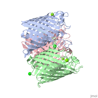
<Structure load='2j1n' size='350' frame='true' align='right' caption='E. coli OmpC' /> | <Structure load='2j1n' size='350' frame='true' align='right' caption='E. coli OmpC' /> | ||
==Porins Overview== | ==Porins Overview== | ||
| - | Porins such as | + | Porins such as OmpC<ref> http://www.proteopedia.org/wiki/index.php/Osmoporin_OmpC_%28E._coli%29 access 12/9/15 </ref>, are proteins located in the outer membrane of gram-negative bacteria or the outer membrane of mitochondria in eukaryotes and function as simple diffusion channels. The simple diffusion channels allow diffusion of sugars, ions, and amino acids to cross the outer membrane. Formation of porins occurs when a signal sequence is used to transfer the porin protein out from the inner membrane of bacteria. For this to occur, chaperone protein Sec B containing an unfolded polypeptide chain sends out a signal. This signal is sent to Sec A ATPase and binds to the Sec B complex. The polypeptide is then released and sent to the Sec E – Sec Y translocation channel where it begins to fold into partially assembled beta-sheets. The lipid-binding region located in the hydrophobic core of the partially assembled beta-sheets will then pull the complex into the outer membrane. Once in the outer membrane, the beta-sheets completely form by the alternating polar/non-polar side chains. Once porins have formed, structural components, functions, and energetic processes can be observed. |
| Line 11: | Line 11: | ||
Porins have many functions in context of its structural components. The main function of porins is passive diffusion channels so that sugars, ions, and amino acids may pass through the outer membrane. For this to occur however, several other functions from other components must occur. One of these components are the hydrophilic chains that line the inside of the beta-barrel of porins. The function of the hydrophilic chains is to create a polar environment within the ring to help sugars, ions, and amino acids to pass through. Another important component is the eyelet or constriction zone located inside the middle of the beta-barrel of porins. The eyelet blocks the water-filled channel of porins and determines what the size capacity and ion selectivity is for a solute to pass through. If a porin has salt-bridges between its N-terminus and C-terminus then it will create more stability for the structure. Lastly, the outside loops formed by the anti-parallel beta-sheets are rigid and packed close together. This allows porins to become highly resistant to proteases and detergents. | Porins have many functions in context of its structural components. The main function of porins is passive diffusion channels so that sugars, ions, and amino acids may pass through the outer membrane. For this to occur however, several other functions from other components must occur. One of these components are the hydrophilic chains that line the inside of the beta-barrel of porins. The function of the hydrophilic chains is to create a polar environment within the ring to help sugars, ions, and amino acids to pass through. Another important component is the eyelet or constriction zone located inside the middle of the beta-barrel of porins. The eyelet blocks the water-filled channel of porins and determines what the size capacity and ion selectivity is for a solute to pass through. If a porin has salt-bridges between its N-terminus and C-terminus then it will create more stability for the structure. Lastly, the outside loops formed by the anti-parallel beta-sheets are rigid and packed close together. This allows porins to become highly resistant to proteases and detergents. | ||
== Energetics == | == Energetics == | ||
| - | The components and functions of porins can be brought together with its energetics to explain porins main function, simple diffusion. [[Image:Facilitated Diffusion.png | thumb | Facilitated Diffusion <ref> “What are the Different Modes of Biotransportation of Drugs – How Drugs Move Across the Cell Membrane?” HubPages. 28 Sept. 2015. Web. 4 Nov. 2015. http://hubpages.com/education/Different-modes-of-biotransport-of-drugs </ref>]] Porins have passive-mediated transport, which is also called facilitated diffusion<ref> Casiday, Rachel. Frey, Regina. “Transport Across Membranes: Energetics and Pumps/Channels.” Academy of Gifted Learners. Department of Chemistry, Washington University. Web. 4 Nov. 2015. https://academyofgiftedlearners.files.wordpress.com/2014/11/chapter13_f141.pdf | + | The components and functions of porins can be brought together with its energetics to explain porins main function, simple diffusion. [[Image:Facilitated Diffusion.png | thumb | Facilitated Diffusion <ref> “What are the Different Modes of Biotransportation of Drugs – How Drugs Move Across the Cell Membrane?” HubPages. 28 Sept. 2015. Web. 4 Nov. 2015. http://hubpages.com/education/Different-modes-of-biotransport-of-drugs </ref>]] Porins have passive-mediated transport, which is also called facilitated diffusion<ref> Casiday, Rachel. Frey, Regina. “Transport Across Membranes: Energetics and Pumps/Channels.” Academy of Gifted Learners. Department of Chemistry, Washington University. Web. 4 Nov. 2015. https://academyofgiftedlearners.files.wordpress.com/2014/11/chapter13_f141.pdf</ref>. Facilitated diffusion allows sugars, ions, amino acids, and polar molecules to pass through the outer membrane. This occurs because of the result of a concentration gradient where solutes travel from an area of high concentration to an area of low concentration. More specifically, porins are channel-forming ionophores which are solvent-filled channels where selected ions can pass through. The selectivity comes from the electrochemical gradient that is formed inside the middle of the porin at the constriction zone. The constriction zone is the eyelet and is surrounded by the positively charged and negatively charged amino acids attached to opposite walls of the beta barrel and the calcium ions. The source of energy for porins and its facilitated diffusion is through its exergonic process. The driving force of this energy is from the concentration gradient, which means no energy is given by transporters. When solutes diffuse through porins from high concentration to low concentration, the entropy becomes positive and creates more disorder and free energy becomes negative. As a result, energy is released. |
| - | </ref>. Facilitated diffusion allows sugars, ions, amino acids, and polar molecules to pass through the outer membrane. This occurs because of the result of a concentration gradient where solutes travel from an area of high concentration to an area of low concentration. More specifically, porins are channel-forming ionophores which are solvent-filled channels where selected ions can pass through. The selectivity comes from the electrochemical gradient that is formed inside the middle of the porin at the constriction zone. The constriction zone is the eyelet and is surrounded by the positively charged and negatively charged amino acids attached to opposite walls of the beta barrel and the calcium ions. The source of energy for porins and its facilitated diffusion is through its exergonic process. The driving force of this energy is from the concentration gradient, which means no energy is given by transporters. When solutes diffuse through porins from high concentration to low concentration, the entropy becomes positive and creates more disorder and free energy becomes negative. As a result, energy is released. | + | |
| - | ==OmpF: | + | ==OmpF:4GCP== |
| - | Certain porins such as 4gcp contain ligands, which operate as it's eyelet. The ligand for 4gcp is an antibody, ampicillin, which is zwitterionic. [[Image:4gcp.png | thumb | OmpF: 4gcp]] | + | Certain porins such as 4gcp<ref> Ziervogel, B.K., Roux, B. “4GCP.” RCSB PDB Protein Data Bank. 6 Feb. 2013. Web. 28 Nov. 2015. http://www.rcsb.org/pdb/explore.do?structureId=4GCP</ref> contain ligands, which operate as it's eyelet. The ligand for 4gcp is an antibody, ampicillin, which is zwitterionic. [[Image:4gcp.png | thumb | OmpF: 4gcp]] |
[[Image:Ampicillin.png | thumb | Ampicillin <ref>http://www.ncbi.nlm.nih.gov/pmc/articles/PMC3545085/figure/F1/ access 12/9/15 </ref>]] The zwitterionic ampicillin antibody contains both positive and negative charge which allows molecules of various charges to transfer through. Other features of the 4gcp porin is that they are found in E.coli, they are non-specific, and they contain two chains (A, B). The rest of the features of the 4gcp porin are the same as the general structure of porins described earlier. | [[Image:Ampicillin.png | thumb | Ampicillin <ref>http://www.ncbi.nlm.nih.gov/pmc/articles/PMC3545085/figure/F1/ access 12/9/15 </ref>]] The zwitterionic ampicillin antibody contains both positive and negative charge which allows molecules of various charges to transfer through. Other features of the 4gcp porin is that they are found in E.coli, they are non-specific, and they contain two chains (A, B). The rest of the features of the 4gcp porin are the same as the general structure of porins described earlier. | ||
| Line 32: | Line 31: | ||
| - | ==PhoE: | + | ==PhoE:1PHO== |
| - | Unlike the 4gcp porin, some porins contain no ligands to operate as it's eyelet. An example of a porin without a ligand would be the 1pho porin. [[Image:1pho.png | thumb | PhoE: 1pho]] Instead of having a ligand as it's eyelet, 1pho has a loop inside, which operates as it's constriction zone or eyelet. This loop is slightly anion selective and creates ion selectivity for molecules to pass through the porin. Other features of the 1pho porin is that they are found in E. coli, they are transports phosphate compounds, and they contain only one chain (A). The rest of the features of the 1pho porin are the same as the general structure of porins described earlier. | + | Unlike the 4gcp porin, some porins contain no ligands to operate as it's eyelet. An example of a porin without a ligand would be the 1pho<ref>Schirmer, T., Cowan, S.W., Jansonius, J.N. “1PHO.” RCSB PDB Protein Data Bank. 13 Jul. 2011. Web. 28 Nov. 2015. http://www.rcsb.org/pdb/explore/explore.do?structureId=1pho</ref> porin. [[Image:1pho.png | thumb | PhoE: 1pho]] Instead of having a ligand as it's eyelet, 1pho has a loop inside, which operates as it's constriction zone or eyelet. This loop is slightly anion selective and creates ion selectivity for molecules to pass through the porin. Other features of the 1pho porin is that they are found in E. coli, they are transports phosphate compounds, and they contain only one chain (A). The rest of the features of the 1pho porin are the same as the general structure of porins described earlier. |
Revision as of 01:24, 17 December 2015
This page is setup for Matt to build his senior project for OU CHEM 4923
|
Contents |
Porins Overview
Porins such as OmpC[1], are proteins located in the outer membrane of gram-negative bacteria or the outer membrane of mitochondria in eukaryotes and function as simple diffusion channels. The simple diffusion channels allow diffusion of sugars, ions, and amino acids to cross the outer membrane. Formation of porins occurs when a signal sequence is used to transfer the porin protein out from the inner membrane of bacteria. For this to occur, chaperone protein Sec B containing an unfolded polypeptide chain sends out a signal. This signal is sent to Sec A ATPase and binds to the Sec B complex. The polypeptide is then released and sent to the Sec E – Sec Y translocation channel where it begins to fold into partially assembled beta-sheets. The lipid-binding region located in the hydrophobic core of the partially assembled beta-sheets will then pull the complex into the outer membrane. Once in the outer membrane, the beta-sheets completely form by the alternating polar/non-polar side chains. Once porins have formed, structural components, functions, and energetic processes can be observed.
Structure
The general structure of porins is most recognized by its secondary structures.
Function
Porins have many functions in context of its structural components. The main function of porins is passive diffusion channels so that sugars, ions, and amino acids may pass through the outer membrane. For this to occur however, several other functions from other components must occur. One of these components are the hydrophilic chains that line the inside of the beta-barrel of porins. The function of the hydrophilic chains is to create a polar environment within the ring to help sugars, ions, and amino acids to pass through. Another important component is the eyelet or constriction zone located inside the middle of the beta-barrel of porins. The eyelet blocks the water-filled channel of porins and determines what the size capacity and ion selectivity is for a solute to pass through. If a porin has salt-bridges between its N-terminus and C-terminus then it will create more stability for the structure. Lastly, the outside loops formed by the anti-parallel beta-sheets are rigid and packed close together. This allows porins to become highly resistant to proteases and detergents.
Energetics
The components and functions of porins can be brought together with its energetics to explain porins main function, simple diffusion.
OmpF:4GCP
Certain porins such as 4gcp[5] contain ligands, which operate as it's eyelet. The ligand for 4gcp is an antibody, ampicillin, which is zwitterionic.

PhoE:1PHO
Unlike the 4gcp porin, some porins contain no ligands to operate as it's eyelet. An example of a porin without a ligand would be the 1pho[7] porin.
References
- ↑ http://www.proteopedia.org/wiki/index.php/Osmoporin_OmpC_%28E._coli%29 access 12/9/15
- ↑ “Porin Channel Protein in Escherichia coli Outer Membrane.” Rs.Noda. Web. 4 Nov. 2015. http://www.rs.noda.tus.ac.jp/~biost/OPFU/yama/public_html/study/folding/md/porin/porin.htm
- ↑ “What are the Different Modes of Biotransportation of Drugs – How Drugs Move Across the Cell Membrane?” HubPages. 28 Sept. 2015. Web. 4 Nov. 2015. http://hubpages.com/education/Different-modes-of-biotransport-of-drugs
- ↑ Casiday, Rachel. Frey, Regina. “Transport Across Membranes: Energetics and Pumps/Channels.” Academy of Gifted Learners. Department of Chemistry, Washington University. Web. 4 Nov. 2015. https://academyofgiftedlearners.files.wordpress.com/2014/11/chapter13_f141.pdf
- ↑ Ziervogel, B.K., Roux, B. “4GCP.” RCSB PDB Protein Data Bank. 6 Feb. 2013. Web. 28 Nov. 2015. http://www.rcsb.org/pdb/explore.do?structureId=4GCP
- ↑ http://www.ncbi.nlm.nih.gov/pmc/articles/PMC3545085/figure/F1/ access 12/9/15
- ↑ Schirmer, T., Cowan, S.W., Jansonius, J.N. “1PHO.” RCSB PDB Protein Data Bank. 13 Jul. 2011. Web. 28 Nov. 2015. http://www.rcsb.org/pdb/explore/explore.do?structureId=1pho
[1]
Two in-text citations from same source:
First time it's referenced: [2]
Subsequent times: Cite error: Invalid <ref> tag;
refs with no name must have content
