PDZ and LIM domain protein
From Proteopedia
(Difference between revisions)
| Line 1: | Line 1: | ||
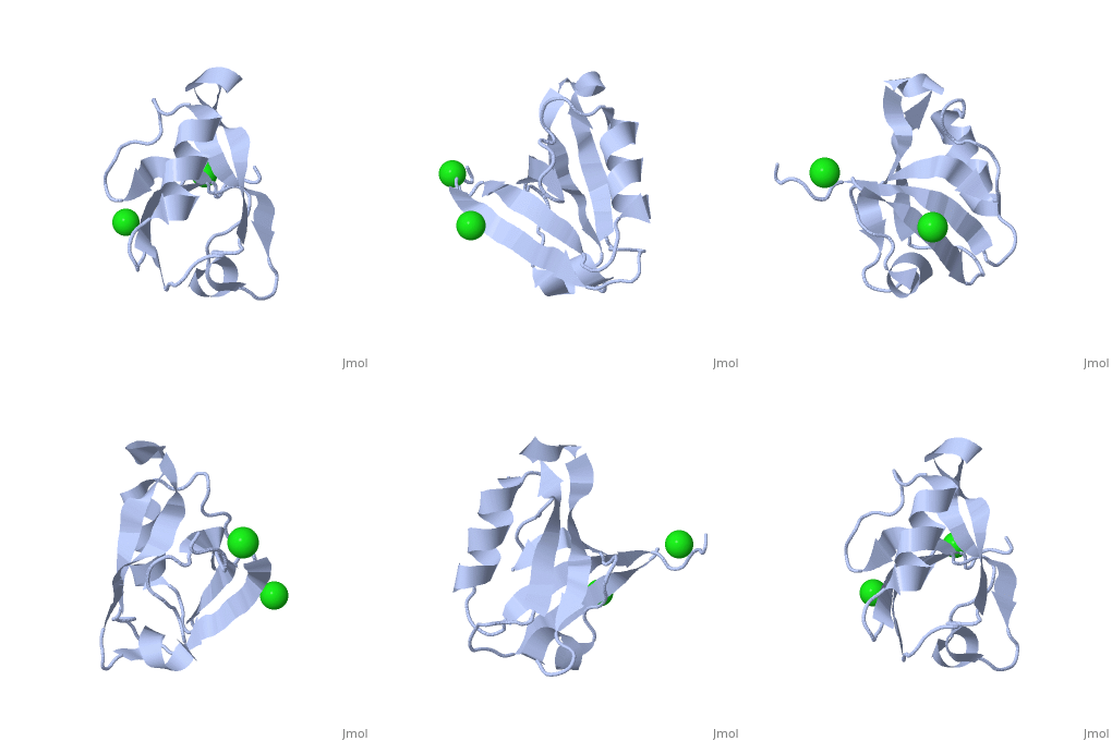
<StructureSection load='2pa1' size='350' side='right' caption='Structure of PDZ and LIM domain protein 2 PDZ domain with C-terminal of β-tropomyosin and Cl- ions (PDB entry [[2pa1]])' scene=''> | <StructureSection load='2pa1' size='350' side='right' caption='Structure of PDZ and LIM domain protein 2 PDZ domain with C-terminal of β-tropomyosin and Cl- ions (PDB entry [[2pa1]])' scene=''> | ||
| - | '''PDZ and LIM domain proteins''' (PDLIM) contain PDZ and LIM domains. PDLIM binds α-actinin via its PDZ domain and other proteins via its LIM domain. PDLIM is implicated in cardiac and skeletal muscle structure. For details on PDLIM1 see [[Group:MUZIC:ALP]]. | + | '''PDZ and LIM domain proteins''' (PDLIM) contain PDZ and LIM domains. PDLIM binds α-actinin via its PDZ domain and other proteins via its LIM domain. PDLIM is implicated in cardiac and skeletal muscle structure.<br /> For details on PDLIM1 see [[Group:MUZIC:ALP]].<br />For details on PDLIM see [[Group:MUZIC:ZASP]]. |
</StructureSection> | </StructureSection> | ||
| Line 40: | Line 40: | ||
**[[2q3g]] – hPDLIM PDZ domain + β-tropomysin C terminal <br /> | **[[2q3g]] – hPDLIM PDZ domain + β-tropomysin C terminal <br /> | ||
| + | |||
| + | *PDLIM | ||
| + | |||
| + | **[[1rgw]] – hPDLIM PDZ domain - NMR<br /> | ||
}} | }} | ||
[[Category:Topic Page]] | [[Category:Topic Page]] | ||
Revision as of 11:43, 6 January 2016
| |||||||||||
3D structures of PDZ and LIM domain protein
Updated on 06-January-2016

