This old version of Proteopedia is provided for student assignments while the new version is undergoing repairs. Content and edits done in this old version of Proteopedia after March 1, 2026 will eventually be lost when it is retired in about June of 2026.
Apply for new accounts at the new Proteopedia. Your logins will work in both the old and new versions.
Thrombin
From Proteopedia
(Difference between revisions)
| Line 25: | Line 25: | ||
By balancing substrate specificity, activity, and inhibition thrombin plays a central role in the blood coagulation cascade. <ref name="three"/> | By balancing substrate specificity, activity, and inhibition thrombin plays a central role in the blood coagulation cascade. <ref name="three"/> | ||
[[Image:Substrates.png|300px|center|thumb| Coagulation related substrates of thrombin, excluding serpin inhibitors.]] | [[Image:Substrates.png|300px|center|thumb| Coagulation related substrates of thrombin, excluding serpin inhibitors.]] | ||
| - | + | {{Clear}} | |
==The Thrombin Life Cycle== | ==The Thrombin Life Cycle== | ||
Upon tissue damage [http://en.wikipedia.org/wiki/Tissue_factor tissue factor (TF)] is released by subendothelial cells. This interacts with circulating [[Factor_VII| FVIIa]], a zymogen-like serine protease, significantly increasing its activity. '''The FVIIa-TF complex (extrinsic Xase) activates FVII, FIX, and FX'''. The now active '''FXa cleaves prothrombin''' bound to membranes through its [http://en.wikipedia.org/wiki/Gla_domain gamma-carboxyglutamyl (Gla) domain], activating it to thrombin. | Upon tissue damage [http://en.wikipedia.org/wiki/Tissue_factor tissue factor (TF)] is released by subendothelial cells. This interacts with circulating [[Factor_VII| FVIIa]], a zymogen-like serine protease, significantly increasing its activity. '''The FVIIa-TF complex (extrinsic Xase) activates FVII, FIX, and FX'''. The now active '''FXa cleaves prothrombin''' bound to membranes through its [http://en.wikipedia.org/wiki/Gla_domain gamma-carboxyglutamyl (Gla) domain], activating it to thrombin. | ||
| Line 41: | Line 41: | ||
==Prothrombin Activation== | ==Prothrombin Activation== | ||
| - | [[Image:Prothrombin activation scheme_3.png|300px| | + | [[Image:Prothrombin activation scheme_3.png|300px|left|thumb| Activation scheme of in vivo activation of prothrombin by FXa in the absence of FVa.]] |
| - | [[Image:Prothrombin activation scheme_nofva3.png|300px| | + | {{Clear}} |
| + | [[Image:Prothrombin activation scheme_nofva3.png|300px|left|thumb| Activation scheme of in vivo activation of prothrombin by the prothrombinase complex in presence of calcium and a phospholipid bilayer]] | ||
| + | {{Clear}} | ||
Prothrombin is the zymogen form of thrombin. From N-terminal to C-terminal it consists of a Gla domain, two kringle domains, and a catalytic domain. The Gla domain is formed by vitamin K dependent carboxylation of glutamate residues.<ref>PMID: 18374193</ref> | Prothrombin is the zymogen form of thrombin. From N-terminal to C-terminal it consists of a Gla domain, two kringle domains, and a catalytic domain. The Gla domain is formed by vitamin K dependent carboxylation of glutamate residues.<ref>PMID: 18374193</ref> | ||
| Line 53: | Line 55: | ||
[[Image:Electrostatic labeled.png|300px|right|thumb| Thrombin (1PPB) overlayed with electrostatic surface. Structural features 60-loop, γ-loop, exosite I, and exosite II labeled]] | [[Image:Electrostatic labeled.png|300px|right|thumb| Thrombin (1PPB) overlayed with electrostatic surface. Structural features 60-loop, γ-loop, exosite I, and exosite II labeled]] | ||
| + | {{Clear}} | ||
Thrombin is a α/β heterodimer composed of a 36 amino acid A chain and 259 amino acid B chain connected by a <scene name='58/583418/Disulfides_nospin/1'>disufide</scene> bridge between Cys1 and Cys122, in addition to 3 other intrachain disulfide bonds.<ref name='eight'>PMID: 2583108</ref> Its overall fold is similar to trypsin and chymotrypsin and it belongs to the [http://merops.sanger.ac.uk/cgi-bin/famsum?family=s1 peptidase S1 protease family]<ref>PMID: 18768474</ref>. It is an overall spherical protein with approximate dimensions of 45 Å X 45 Å X 50 Å.<ref name='eight'/> | Thrombin is a α/β heterodimer composed of a 36 amino acid A chain and 259 amino acid B chain connected by a <scene name='58/583418/Disulfides_nospin/1'>disufide</scene> bridge between Cys1 and Cys122, in addition to 3 other intrachain disulfide bonds.<ref name='eight'>PMID: 2583108</ref> Its overall fold is similar to trypsin and chymotrypsin and it belongs to the [http://merops.sanger.ac.uk/cgi-bin/famsum?family=s1 peptidase S1 protease family]<ref>PMID: 18768474</ref>. It is an overall spherical protein with approximate dimensions of 45 Å X 45 Å X 50 Å.<ref name='eight'/> | ||
Important structural features include: | Important structural features include: | ||
| Line 79: | Line 82: | ||
The B chain consists of <scene name='Serine_Protease/Domains/1'>two domains</scene>. As is true for all of the "trypsin-like" serine proteases, each of the two thrombin domains consists mainly of a 6-stranded, antiparallel beta barrel. The specificity pocket (here filled with the Lys sidechain of the PPACK inhibitor) is in one side of the throat of the domain 2beta barrel, and the activation site is close next to it. | The B chain consists of <scene name='Serine_Protease/Domains/1'>two domains</scene>. As is true for all of the "trypsin-like" serine proteases, each of the two thrombin domains consists mainly of a 6-stranded, antiparallel beta barrel. The specificity pocket (here filled with the Lys sidechain of the PPACK inhibitor) is in one side of the throat of the domain 2beta barrel, and the activation site is close next to it. | ||
| - | + | ||
==Allostery== | ==Allostery== | ||
| Line 102: | Line 105: | ||
[[Image:Capping.png|450px|center|thumb| “Capping box” motif in alpha thrombin (PDB: 1PPB) represented by His230 (Ncap) side chain and main chain hydrogen bonded with the backbone nitrogen of Arg233 (N3). An additional feature is a weak hydrophobic interaction between Thr229 (N’) and Val234 (N4) termed the “hydrophobic staple.” This motif derives it’s name from the box shaped hydrogen bonding pattern.]] | [[Image:Capping.png|450px|center|thumb| “Capping box” motif in alpha thrombin (PDB: 1PPB) represented by His230 (Ncap) side chain and main chain hydrogen bonded with the backbone nitrogen of Arg233 (N3). An additional feature is a weak hydrophobic interaction between Thr229 (N’) and Val234 (N4) termed the “hydrophobic staple.” This motif derives it’s name from the box shaped hydrogen bonding pattern.]] | ||
| - | + | {{Clear}} | |
[[Image:Cation pi.png|450px|center|thumb| A cation-π interaction between Trp128 and Arg 129 in alpha thrombin (PDB: 2BDY). The guanidinium carbon is 3.6 angstroms from the top edge of the Trp. It is expected that the epsilon nitrogen forms the primary cation-π interaction. The electrostatic and Van der Waals interaction energies were calculated to be -3.29 kcal/mol and -3.08 kcal/mol respectively by the CaPTURE program (http://capture.caltech.edu/result.cgi).]] | [[Image:Cation pi.png|450px|center|thumb| A cation-π interaction between Trp128 and Arg 129 in alpha thrombin (PDB: 2BDY). The guanidinium carbon is 3.6 angstroms from the top edge of the Trp. It is expected that the epsilon nitrogen forms the primary cation-π interaction. The electrostatic and Van der Waals interaction energies were calculated to be -3.29 kcal/mol and -3.08 kcal/mol respectively by the CaPTURE program (http://capture.caltech.edu/result.cgi).]] | ||
| + | {{Clear}} | ||
</StructureSection> | </StructureSection> | ||
Revision as of 07:56, 25 January 2016
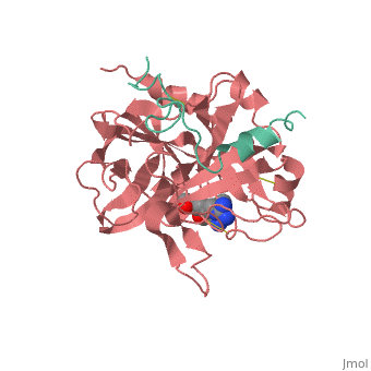
| |||||||||||


3D Structures of thrombin
Updated on 25-January-2016
References
- ↑ Fenton JW 2nd. Thrombin specificity. Ann N Y Acad Sci. 1981;370:468-95. PMID:7023326
- ↑ 2.0 2.1 Coughlin SR. Thrombin signalling and protease-activated receptors. Nature. 2000 Sep 14;407(6801):258-64. PMID:11001069 doi:http://dx.doi.org/10.1038/35025229
- ↑ Crawley JT, Lam JK, Rance JB, Mollica LR, O'Donnell JS, Lane DA. Proteolytic inactivation of ADAMTS13 by thrombin and plasmin. Blood. 2005 Feb 1;105(3):1085-93. Epub 2004 Sep 23. PMID:15388580 doi:http://dx.doi.org/10.1182/blood-2004-03-1101
- ↑ 4.0 4.1 4.2 4.3 4.4 4.5 4.6 Lane DA, Philippou H, Huntington JA. Directing thrombin. Blood. 2005 Oct 15;106(8):2605-12. Epub 2005 Jun 30. PMID:15994286 doi:http://dx.doi.org/10.1182/blood-2005-04-1710
- ↑ Takagi T, Doolittle RF. Amino acid sequence studies on factor XIII and the peptide released during its activation by thrombin. Biochemistry. 1974 Feb 12;13(4):750-6. PMID:4811064
- ↑ Miljic P, Heylen E, Willemse J, Djordjevic V, Radojkovic D, Colovic M, Elezovic I, Hendriks D. Thrombin activatable fibrinolysis inhibitor (TAFI): a molecular link between coagulation and fibrinolysis. Srp Arh Celok Lek. 2010 Jan;138 Suppl 1:74-8. PMID:20229688
- ↑ 7.0 7.1 7.2 7.3 Huntington JA. Natural inhibitors of thrombin. Thromb Haemost. 2014 Apr 1;111(4):583-9. doi: 10.1160/TH13-10-0811. Epub 2014 Jan, 30. PMID:24477356 doi:http://dx.doi.org/10.1160/TH13-10-0811
- ↑ 8.0 8.1 8.2 8.3 8.4 Huntington JA. Thrombin inhibition by the serpins. J Thromb Haemost. 2013 Jun;11 Suppl 1:254-64. doi: 10.1111/jth.12252. PMID:23809129 doi:http://dx.doi.org/10.1111/jth.12252
- ↑ Esmon CT. The regulation of natural anticoagulant pathways. Science. 1987 Mar 13;235(4794):1348-52. PMID:3029867
- ↑ Kalafatis M, Rand MD, Mann KG. The mechanism of inactivation of human factor V and human factor Va by activated protein C. J Biol Chem. 1994 Dec 16;269(50):31869-80. PMID:7989361
- ↑ 11.0 11.1 Lu D, Kalafatis M, Mann KG, Long GL. Comparison of activated protein C/protein S-mediated inactivation of human factor VIII and factor V. Blood. 1996 Jun 1;87(11):4708-17. PMID:8639840
- ↑ Duga S, Asselta R, Tenchini ML. Coagulation factor V. Int J Biochem Cell Biol. 2004 Aug;36(8):1393-9. PMID:15147718 doi:http://dx.doi.org/10.1016/j.biocel.2003.08.002
- ↑ Saenko EL, Shima M, Sarafanov AG. Role of activation of the coagulation factor VIII in interaction with vWf, phospholipid, and functioning within the factor Xase complex. Trends Cardiovasc Med. 1999 Oct;9(7):185-92. PMID:10881749
- ↑ Camire, R. M. (2010). Platelet factor V to the rescue. Blood, 115(4), 753-754. DOI: 10.1182/blood-2009-11-252619
- ↑ Berkner KL. Vitamin K-dependent carboxylation. Vitam Horm. 2008;78:131-56. doi: 10.1016/S0083-6729(07)00007-6. PMID:18374193 doi:http://dx.doi.org/10.1016/S0083-6729(07)00007-6
- ↑ 16.0 16.1 16.2 16.3 16.4 16.5 16.6 16.7 Lechtenberg BC, Freund SM, Huntington JA. An ensemble view of thrombin allostery. Biol Chem. 2012 Sep;393(9):889-98. doi: 10.1515/hsz-2012-0178. PMID:22944689 doi:http://dx.doi.org/10.1515/hsz-2012-0178
- ↑ Tijburg PN, van Heerde WL, Leenhouts HM, Hessing M, Bouma BN, de Groot PG. Formation of meizothrombin as intermediate in factor Xa-catalyzed prothrombin activation on endothelial cells. The influence of thrombin on the reaction mechanism. J Biol Chem. 1991 Feb 25;266(6):4017-22. PMID:1995649
- ↑ Bobofchak KM, Pineda AO, Mathews FS, Di Cera E. Energetic and structural consequences of perturbing Gly-193 in the oxyanion hole of serine proteases. J Biol Chem. 2005 Jul 8;280(27):25644-50. Epub 2005 May 12. PMID:15890651 doi:http://dx.doi.org/10.1074/jbc.M503499200
- ↑ 19.0 19.1 19.2 19.3 Bode W, Mayr I, Baumann U, Huber R, Stone SR, Hofsteenge J. The refined 1.9 A crystal structure of human alpha-thrombin: interaction with D-Phe-Pro-Arg chloromethylketone and significance of the Tyr-Pro-Pro-Trp insertion segment. EMBO J. 1989 Nov;8(11):3467-75. PMID:2583108
- ↑ Page MJ, Di Cera E. Evolution of peptidase diversity. J Biol Chem. 2008 Oct 31;283(44):30010-4. doi: 10.1074/jbc.M804650200. Epub 2008 , Sep 3. PMID:18768474 doi:http://dx.doi.org/10.1074/jbc.M804650200
- ↑ Schechter I, Berger A. On the size of the active site in proteases. I. Papain. 1967. Biochem Biophys Res Commun. 2012 Aug 31;425(3):497-502. doi:, 10.1016/j.bbrc.2012.08.015. PMID:22925665 doi:http://dx.doi.org/10.1016/j.bbrc.2012.08.015
- ↑ Huntington JA. Molecular recognition mechanisms of thrombin. J Thromb Haemost. 2005 Aug;3(8):1861-72. PMID:16102053 doi:http://dx.doi.org/10.1111/j.1538-7836.2005.01363.x
- ↑ Zhang E, Tulinsky A. The molecular environment of the Na+ binding site of thrombin. Biophys Chem. 1997 Jan 31;63(2-3):185-200. PMID:9108691
- ↑ Li W, Johnson DJ, Esmon CT, Huntington JA. Structure of the antithrombin-thrombin-heparin ternary complex reveals the antithrombotic mechanism of heparin. Nat Struct Mol Biol. 2004 Sep;11(9):857-62. Epub 2004 Aug 15. PMID:15311269 doi:10.1038/nsmb811
- ↑ Spronk HM, Borissoff JI, ten Cate H. New insights into modulation of thrombin formation. Curr Atheroscler Rep. 2013 Nov;15(11):363. doi: 10.1007/s11883-013-0363-3. PMID:24026641 doi:http://dx.doi.org/10.1007/s11883-013-0363-3
With participation by User:Cody Couperus
Proteopedia Page Contributors and Editors (what is this?)
Michal Harel, Alexander Berchansky, Cody Couperus, Joel L. Sussman
