We apologize for Proteopedia being slow to respond. For the past two years, a new implementation of Proteopedia has been being built. Soon, it will replace this 18-year old system. All existing content will be moved to the new system at a date that will be announced here.
Sandbox Reserved 1125
From Proteopedia
(Difference between revisions)
| Line 71: | Line 71: | ||
== Regulation by inhibitors == | == Regulation by inhibitors == | ||
Petites info Chrichri: | Petites info Chrichri: | ||
| - | <font=MMP activity may subsequently be regulated by the action of inhibitors, notably the tissue inhibitors of MMPs (TIMPs) - TIMP-1, TIMP-2, TIMP-3 and TIMP-4 - and the serum panproteinase inhibitor α2 macroglobulin (Baker et al., 2002) The TIMPs are six-loop disulphide-bonded proteins forming two domains. They interact via their N-terminal three disulphide-bonded loops with the active site cleft of the catalytic domain, although significant interactions of the hemopexin-like domains of MMP-2 and MMP-9 with the C-terminal domains of TIMPs appear to have specific biological relevance. The other MMP domains have distinct functions, such as as exosites for substrate interactions, e.g. the hemopexin-like domains of MMP-1, MMP-8, MMP-13, MMP-14, MMP-16 and MMP-18 are essential for their ability to cleave fibrillar collagens and the fibronectin-like domains of MMP-2 and MMP-9 confer their binding to denatured collagen substrates. The hemopexin-like domain of MMP-14 can homodimerise in order to promote its clustering at the cell surface, a property that promotes its activity. The hemopexin-like domain confers the ability to interact with other extracellular matrix components and cell adhesion molecules and may be of significance in the determination of specific pericellular locations of individual MMPs. | + | <font color='red'>MMP activity may subsequently be regulated by the action of inhibitors, notably the tissue inhibitors of MMPs (TIMPs) - TIMP-1, TIMP-2, TIMP-3 and TIMP-4 - and the serum panproteinase inhibitor α2 macroglobulin (Baker et al., 2002) The TIMPs are six-loop disulphide-bonded proteins forming two domains. They interact via their N-terminal three disulphide-bonded loops with the active site cleft of the catalytic domain, although significant interactions of the hemopexin-like domains of MMP-2 and MMP-9 with the C-terminal domains of TIMPs appear to have specific biological relevance. The other MMP domains have distinct functions, such as as exosites for substrate interactions, e.g. the hemopexin-like domains of MMP-1, MMP-8, MMP-13, MMP-14, MMP-16 and MMP-18 are essential for their ability to cleave fibrillar collagens and the fibronectin-like domains of MMP-2 and MMP-9 confer their binding to denatured collagen substrates. The hemopexin-like domain of MMP-14 can homodimerise in order to promote its clustering at the cell surface, a property that promotes its activity. The hemopexin-like domain confers the ability to interact with other extracellular matrix components and cell adhesion molecules and may be of significance in the determination of specific pericellular locations of individual MMPs. |
| - | + | ||
The MMPs are regulated at the transcriptional and post-transcriptional levels, as well as by activation, inhibition and cell/ECM localization, which allows tissue-specific spatial and temporal patterns of functional activity. Expression levels may be modulated by different cytokines, growth factors, hormones, extracellular matrix interactions and cytoskeletal changes through specific elements in the MMP promoters governing transcriptional regulation. Sequestration of the secreted MMPs in Golgi vesicles has been described for many stimulated cells, as has storage of MMP-8 and MMP-9 in the secretory granules of PMN leucocytes. The membrane-associated MMPs appear to have distinct trafficking pathways to specific sites at the cell surface. Association of some MMPs with integrins and other cell surface receptors has been described, e.g. MMP-1-integrin-α2β1, MMP-2-integrin-αVβ3, MMP-14-integrin-α2β1/αVβ3, MMP-7-CD44 and MMP-9-CD44. Many MMPs bind to specific ECM components (see above). With the exception of very rapidly remodeling tissues, extracellular levels of MMPs tend to be quite low, and unambiguous immunohistochemical detection is challenging. | The MMPs are regulated at the transcriptional and post-transcriptional levels, as well as by activation, inhibition and cell/ECM localization, which allows tissue-specific spatial and temporal patterns of functional activity. Expression levels may be modulated by different cytokines, growth factors, hormones, extracellular matrix interactions and cytoskeletal changes through specific elements in the MMP promoters governing transcriptional regulation. Sequestration of the secreted MMPs in Golgi vesicles has been described for many stimulated cells, as has storage of MMP-8 and MMP-9 in the secretory granules of PMN leucocytes. The membrane-associated MMPs appear to have distinct trafficking pathways to specific sites at the cell surface. Association of some MMPs with integrins and other cell surface receptors has been described, e.g. MMP-1-integrin-α2β1, MMP-2-integrin-αVβ3, MMP-14-integrin-α2β1/αVβ3, MMP-7-CD44 and MMP-9-CD44. Many MMPs bind to specific ECM components (see above). With the exception of very rapidly remodeling tissues, extracellular levels of MMPs tend to be quite low, and unambiguous immunohistochemical detection is challenging. | ||
| - | + | The four TIMPs act as a further level of extracellular regulation and also have specific patterns of gene regulation and tissue-specific expression. TIMP-3 is unusual in that it is largely sequestered into the extracellular matrix or at the cell surface via heparan sulphate proteoglycans. Individual TIMPs differ in their ability to inhibit different MMPs; TIMP-1 is a poor inhibitor of MMP-14, MMP-16 and MMP-19. In addition there are specific interactions of TIMP-1 with proMMP-9, of TIMP-2 with proMMP-2 and of TIMP-3 with both proMMP-2 and proMMP-9 by binding through their three C-terminal disulphide-bonded loops, which allows complexes of the inactive MMPs to be formed, as well as giving very tight-binding active enzyme complexes. The true significance of this has only been elucidated for proMMP-2, where the TIMP-2 complex allows binding of the MMP to MMP-14 at the cell surface, promoting its activation and potentially focusing proteolysis to specific sites. The activation of proMMPs in general is probably strictly pericellular, e.g. where plasmin, generated by the activity of urokinase-type plasminogen activator, is an initiator of activation cascades. If there is an excess of TIMPs and serine proteinase inhibitors in the environment, these may also confine activity to the local environment. There is a further level of regulation of the MMPs through clearance by endocytosis. Little is known of the fate of most MMP-TIMP complexes, but complexes with α2 macroglobulin are thought to be endocytosed after binding to the low density lipoprotein receptor related protein (LRP). Thrombospondin 2 modulates both MMP-9-TIMP-1 and MMP-2 internalisation via LRP. The membrane-associated proteinase MMP-14 is endocytosed via clathrin- and nonclathrin-mediated pathways and may recycle to the cell surface in some situations. The other MT-MMPs probably have similar properties.</font><ref>PMID:12235282</ref> | |
| - | The four TIMPs act as a further level of extracellular regulation and also have specific patterns of gene regulation and tissue-specific expression. TIMP-3 is unusual in that it is largely sequestered into the extracellular matrix or at the cell surface via heparan sulphate proteoglycans. Individual TIMPs differ in their ability to inhibit different MMPs; TIMP-1 is a poor inhibitor of MMP-14, MMP-16 and MMP-19. In addition there are specific interactions of TIMP-1 with proMMP-9, of TIMP-2 with proMMP-2 and of TIMP-3 with both proMMP-2 and proMMP-9 by binding through their three C-terminal disulphide-bonded loops, which allows complexes of the inactive MMPs to be formed, as well as giving very tight-binding active enzyme complexes. The true significance of this has only been elucidated for proMMP-2, where the TIMP-2 complex allows binding of the MMP to MMP-14 at the cell surface, promoting its activation and potentially focusing proteolysis to specific sites. The activation of proMMPs in general is probably strictly pericellular, e.g. where plasmin, generated by the activity of urokinase-type plasminogen activator, is an initiator of activation cascades. If there is an excess of TIMPs and serine proteinase inhibitors in the environment, these may also confine activity to the local environment. There is a further level of regulation of the MMPs through clearance by endocytosis. Little is known of the fate of most MMP-TIMP complexes, but complexes with α2 macroglobulin are thought to be endocytosed after binding to the low density lipoprotein receptor related protein (LRP). Thrombospondin 2 modulates both MMP-9-TIMP-1 and MMP-2 internalisation via LRP. The membrane-associated proteinase MMP-14 is endocytosed via clathrin- and nonclathrin-mediated pathways and may recycle to the cell surface in some situations. The other MT-MMPs probably have similar properties. | + | |
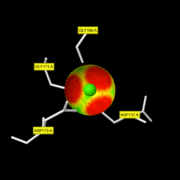
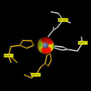
<scene name='71/719866/Timp1/1'>TIMP</scene> | <scene name='71/719866/Timp1/1'>TIMP</scene> | ||
Revision as of 08:21, 29 January 2016
MMP-8
MMP-8, also called, Neutrophil collagenase or Collagenase 2, is a zinc-dependent and calcium-dependent enzyme. It belongs to the matrix metalloproteinase (MMP) family which is involved in the breakdown of extracellular matrix in embryonic development, reproduction, and tissue remodeling, as well as in disease processes, such as arthritis and metastasis. The gene coding this family is localized on the chromosome 11 of Homo sapiens with 467 residues.[1]
| |||||||||||
References
- ↑ "MMP-8 matrix metallopeptidase 8 (neutrophil collagenase)"
- ↑ "Metalloendopeptidase activity"
- ↑ Stams T, Spurlino JC, Smith DL, Wahl RC, Ho TF, Qoronfleh MW, Banks TM, Rubin B. Structure of human neutrophil collagenase reveals large S1' specificity pocket. Nat Struct Biol. 1994 Feb;1(2):119-23. PMID:7656015
- ↑ 4.0 4.1 Substrate specificity of MMPs
- ↑ Bode W, Reinemer P, Huber R, Kleine T, Schnierer S, Tschesche H. The X-ray crystal structure of the catalytic domain of human neutrophil collagenase inhibited by a substrate analogue reveals the essentials for catalysis and specificity. EMBO J. 1994 Mar 15;13(6):1263-9. PMID:8137810
- ↑ Bode W, Reinemer P, Huber R, Kleine T, Schnierer S, Tschesche H. The X-ray crystal structure of the catalytic domain of human neutrophil collagenase inhibited by a substrate analogue reveals the essentials for catalysis and specificity. EMBO J. 1994 Mar 15;13(6):1263-9. PMID:8137810
- ↑ Hirose T, Patterson C, Pourmotabbed T, Mainardi CL, Hasty KA. Structure-function relationship of human neutrophil collagenase: identification of regions responsible for substrate specificity and general proteinase activity. Proc Natl Acad Sci U S A. 1993 Apr 1;90(7):2569-73. PMID:8464863
- ↑ Knauper V, Osthues A, DeClerck YA, Langley KE, Blaser J, Tschesche H. Fragmentation of human polymorphonuclear-leucocyte collagenase. Biochem J. 1993 May 1;291 ( Pt 3):847-54. PMID:8489511
- ↑ Van Wart HE, Birkedal-Hansen H. The cysteine switch: a principle of regulation of metalloproteinase activity with potential applicability to the entire matrix metalloproteinase gene family. Proc Natl Acad Sci U S A. 1990 Jul;87(14):5578-82. PMID:2164689
- ↑ Welgus HG, Jeffrey JJ, Eisen AZ. Human skin fibroblast collagenase. Assessment of activation energy and deuterium isotope effect with collagenous substrates. J Biol Chem. 1981 Sep 25;256(18):9516-21. PMID:6270090
- ↑ Visse R, Nagase H. Matrix metalloproteinases and tissue inhibitors of metalloproteinases: structure, function, and biochemistry. Circ Res. 2003 May 2;92(8):827-39. PMID:12730128 doi:http://dx.doi.org/10.1161/01.RES.0000070112.80711.3D
- ↑ Knauper V, Docherty AJ, Smith B, Tschesche H, Murphy G. Analysis of the contribution of the hinge region of human neutrophil collagenase (HNC, MMP-8) to stability and collagenolytic activity by alanine scanning mutagenesis. FEBS Lett. 1997 Mar 17;405(1):60-4. PMID:9094424
- ↑ "Neutrophil collagenase"
- ↑ Baker AH, Edwards DR, Murphy G. Metalloproteinase inhibitors: biological actions and therapeutic opportunities. J Cell Sci. 2002 Oct 1;115(Pt 19):3719-27. PMID:12235282
- ↑ "Extra Binding Region Induced by Non-Zinc Chelating Inhibitors into the S1′ Subsite of Matrix Metalloproteinase 8"
- ↑ Balbin M, Fueyo A, Knauper V, Pendas AM, Lopez JM, Jimenez MG, Murphy G, Lopez-Otin C. Collagenase 2 (MMP-8) expression in murine tissue-remodeling processes. Analysis of its potential role in postpartum involution of the uterus. J Biol Chem. 1998 Sep 11;273(37):23959-68. PMID:9727011
RESSOURCE : Image:2oy4 mm1.pdb ( la structure du monomère )


