Classification
EC 3.4.24.34
This classification means that this enzyme:
- is a hydrolase: it hydrolyzes covalent bonds
- is an endopeptidase: it cleaves peptide bond
- cleaves interstitial collagens in the triple helical domain (at a site about three-fourths away from the N-terminus)
The metalloendopeptidase activity is defined by a mechanism in which water acts as a nucleophile, one or two metal ions hold the water molecule in place, and charged amino acid side chains are ligands for the metal ions.[2]
The difference between this classification and EC 3.4.24.7 is that this enzyme cleaves type III collagen more slowly than type I.
(On BRENDA, you can find all informations about the MMP8 enzyme like, for example, a list of different substrates or inhibitors)
To see
[1]
[2]
[3]
[3]
[4] pockets
[5]
Structure and domains
MMP-8 is composed of several domains: a propeptide, a catalytic domain, a hinge region, and a C-terminal hemopexinlike domain.[4].
Propeptide
It corresponds to 79 aminoacids, from Phe21 to Met100.
The sequence of residue is: FPVSSKEKNTKTVQDYLEKFYQLPSNQYQSTRKNGTNVIVEKLKEMQRFFGLNVTGKPNEETLDMMKKPRCGVPDSGGFM
Catalytic domain
Thanks to X-ray crystallography, the catalytic domain structure has been solved with 1,7 Å resolution (2OY4).This domain is composed of 157 residues, from Met86 to Gly242, organized in and .The protein folding and especially the zinc environment of the collagenase catalytic domain is very close to the astacins and the snake venom metalloproteinases. The catalytic domain alone has proteolytic activity against other protein substrates and synthetic substrates.[5]
Ca2+ interactions
This enzyme binds 3 Ca ions, 2 of them in the catalytic domain, which are packed against the top of the beta sheet and mostly have a structural function, stabilizing the catalytic domain.
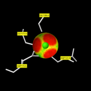
The residues involved in the Ca996 interactions (coordinate bonds) are .
Zn2+ interactions
The zinc-binding motif HEXGHXXGXXH presents in the catalytic domain is characteristic for the protease activity of MMP-8.
Zn999 : the catalytic zinc
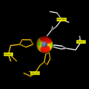
It is involved in the catalytic activity and is situated at the bottom of the active-site. This ion is penta-coordinated with: His197, His201 and His207 of MMP-8 and with the carbonyl and the hydroxyl oxygen of the hydroxamic acid moiety of the inhibitor. This discovery has been made thanks to the Pro-Leu-Gly-hydroxylamine inhibitor.[6] On this you can only see the 3 His of MMP-8 with the Zn999. The fourth ligand of the catalytic zinc is a water molecule.
Zn998 : the structural zinc
The residues involved in the Zn998 interactions are . The glutamic acid adjacent to the first histidine is essential for catalysis. It should be noted that scientists were unable to exchange or remove this Zinc in their crystals, which is suggesting that there is a tight interaction with MMP-8.[7]
Hinge domain
It corresponds to a short linker region from G242 to P258, with the following sequence: GLSSNPIQPTGPSTPKP, between the catalytic and the hemopexin domains. The exact role of this domain isn't very well clear but it's known that autoproteolysis could occurred in MMP8 leading to an unstable protein and different mutants[8] were made in the hinge region and it shown that stability of MMP8 could be increased, decreased or unchanged. Moreover, sequence alignements of collagenolytic MMPs in this hinge domain reveal that they all have the four prolines in the same positions, suggesting that these prolines could be important for the specific collagenolytic activity.
Hemopexin domain
The hemopexin domain has two conserved cysteines that are disulfide bonded. Mutation of those cysteines to alanines [9] or reduction and alkylation destroys collagenolytic activity (K. Suzuki and H.Nagase, unpublished results).[4]
[6]
This domain is localized outside of the catalytic domain shown in this page. It is essential for the substrate recognition of MMP-8 and the single catalytic domain of MMP-8 is not able to cleave collagen. When this hemopexin-like domain is removed, the protein loses its ability to cleave collagen. However, neutrophil collagenase is still able to cleave other substrate.
It seems that the collagen binds to two sites on MMP-8 : one in the catalytic site and another in the hemopexin domain. One hypothesis is that when the collagen binds to both sites, its helical structure is destabilized and unwound. Thus, the cleavage site of collagen is accessible and the cleavage reaction can occur.
Mechanism
MMP-8 is secreted as inactive proproteins and then activated after a cleavage by extracellular proteinases. Indeed, it can't be activated without removal of the activation peptide. After this protease activation, recent evidences suggest that, there is formation of an intramolecular complex between the Cysteine residue (Cys91) of the propeptide domain and essential zinc atom in the catalytic domain. It is called the Cysteine-switch: it inhibits the action of MMP-8. This discovery is unprecedented in enzymology and offers the opportunity for multiple modes of physiological activation of MMP-8. Moreover, since conditions in different cells and tissues may match those necessary to effect one of these activation modes for a given MMP, this may offer metabolic flexibility in the control of MMP activation.[10]
To express collagenolytic activity, MMP-8 needs to have both the catalytic and hemopexin domains. The linker peptide can position the hemopexin domain in such a way that it bends over the active site of the catalytic domain. But understanding how the Hemopexin domain assists in the cleavage of collagen is elusive.[11] Thus, the collagen would be captured between these two domains. However, the active site cannot accommodate the entire triple helix in a native state. The linker peptide would, by means of its collagen-like conformation, change the quaternary structure of the captured collagen. Interactions between proline residues of the collagenase and a specific region of the collagen would generate a “proline zipper,” resulting in destabilization of the cleavage site area of the collagen. After destabilization, one chain of the triple helix fits in the specificity pocket or to the right of the active-site zinc. At first, the Gly residue of the substrate binds the thanks to the Zn2+ atom. When it binds it takes the place of unstable water molecules and establishes stabilizing interactions with the active site thanks to its C terminal part.[12] The carboxyl group of the glutamate serves as a general base to draw a proton from the displaced water molecule, thereby facilitating the nucleophilic attack of the water molecule on the carbonyl carbon of the peptide scissile bond. Then, the Alanine residue of the enzyme makes a hydrogen bond with the NH group of the substrate. Moreover, this NH group becomes the new N-terminus after cleavage.[13]
The cleavage is at Gly775–Ile776 or Leu776 in each alpha-chain of the collagen molecule[14] and takes place at neutral pH. It generates fragments that spontaneously lose their helical conformation, denature to gelatin, and become soluble. The gelatin is then susceptible to attack by gelatinases and other proteases.[15]
Inhibitors of MMP-8
Endogenous inhibitors
The tissue inhibitors of metalloproteinases (TIMPs) are specific inhibitors of the whole family of MMPs proteins. Currently, four TIMPs were identified (TIMP-1, TIMP-2, TIMP-3, TIMP-4).
They are 21 to 29kDa proteins, contain 2 subdomains (N-ter and C-ter) and have a "wedge-like" shape.
This is the N-ter domain which interacts with the catalytic domains of MMPs and impedes their proteolytic activity.[16]
The Cys1 residue is crucial for the inhibitor effect of TIMPs because it can interact with the catalytic zinc of the MMPs. The result is a chelation of the zinc by the N-terminal amino group and the carbonyl group of Cys1.[17] Moreover, this interaction triggers the expulsion of the water molecule which was bound to the zinc.
No structures of MMP-8 with TIMPs are available on PDB. However, the mecanism of inhibition is common to all the MMPs. To see the interaction between the catalytic domain of MMP-3 and TIMP-1 (in green) click .[18]
Synthetic inhibitors
Because endogenous TIMPs have a broad spectrum of action over MMPs, researches were conducted to produce engineered TIMPs and modify their affinity for MMPs. For instance, mutation of the Thr2 of TIMP-1 modify the specificity of this inhibitor as this residue interacts with the S1' pocket of the MMPs.[19]
Besides the modification of TIMPs, the research for MMPs inhibitors resulted in the discovering of molecules with the ability to interact with the catalytic domain of MMPs. The first developped synthetic inhibitors were molecules that mimic the natural substrates of MMPs combined with a zinc chelating groupement.[20]
Numerous range of compounds such as hydroxamate, thiol, pyrimidine and phosphorus based-molecules were developped. Those inhibitors inhibit the activity of MMPs by chelating the catalytic zinc like for MMP-8.
Recently, new range of inhibitors which do not chelate the catalytic zinc were developped. Those compounds target the selectivity regions for substrates of the MMPs rather than binding to the catalytic zinc. For instance, they can interact with the S1' pocket and induce a conformational change like of MMP-8.
Petites info Chrichri:
MMP activity may subsequently be regulated by the action of inhibitors, notably the tissue inhibitors of MMPs (TIMPs) - TIMP-1, TIMP-2, TIMP-3 and TIMP-4 - and the serum panproteinase inhibitor α2 macroglobulin (Baker et al., 2002) The TIMPs are six-loop disulphide-bonded proteins forming two domains. They interact via their N-terminal three disulphide-bonded loops with the active site cleft of the catalytic domain, although significant interactions of the hemopexin-like domains of MMP-2 and MMP-9 with the C-terminal domains of TIMPs appear to have specific biological relevance. The other MMP domains have distinct functions, such as as exosites for substrate interactions, e.g. the hemopexin-like domains of MMP-1, MMP-8, MMP-13, MMP-14, MMP-16 and MMP-18 are essential for their ability to cleave fibrillar collagens and the fibronectin-like domains of MMP-2 and MMP-9 confer their binding to denatured collagen substrates. The hemopexin-like domain of MMP-14 can homodimerise in order to promote its clustering at the cell surface, a property that promotes its activity. The hemopexin-like domain confers the ability to interact with other extracellular matrix components and cell adhesion molecules and may be of significance in the determination of specific pericellular locations of individual MMPs.
The MMPs are regulated at the transcriptional and post-transcriptional levels, as well as by activation, inhibition and cell/ECM localization, which allows tissue-specific spatial and temporal patterns of functional activity. Expression levels may be modulated by different cytokines, growth factors, hormones, extracellular matrix interactions and cytoskeletal changes through specific elements in the MMP promoters governing transcriptional regulation. Sequestration of the secreted MMPs in Golgi vesicles has been described for many stimulated cells, as has storage of MMP-8 and MMP-9 in the secretory granules of PMN leucocytes. The membrane-associated MMPs appear to have distinct trafficking pathways to specific sites at the cell surface. Association of some MMPs with integrins and other cell surface receptors has been described, e.g. MMP-1-integrin-α2β1, MMP-2-integrin-αVβ3, MMP-14-integrin-α2β1/αVβ3, MMP-7-CD44 and MMP-9-CD44. Many MMPs bind to specific ECM components (see above). With the exception of very rapidly remodeling tissues, extracellular levels of MMPs tend to be quite low, and unambiguous immunohistochemical detection is challenging.
The four TIMPs act as a further level of extracellular regulation and also have specific patterns of gene regulation and tissue-specific expression. TIMP-3 is unusual in that it is largely sequestered into the extracellular matrix or at the cell surface via heparan sulphate proteoglycans. Individual TIMPs differ in their ability to inhibit different MMPs; TIMP-1 is a poor inhibitor of MMP-14, MMP-16 and MMP-19. In addition there are specific interactions of TIMP-1 with proMMP-9, of TIMP-2 with proMMP-2 and of TIMP-3 with both proMMP-2 and proMMP-9 by binding through their three C-terminal disulphide-bonded loops, which allows complexes of the inactive MMPs to be formed, as well as giving very tight-binding active enzyme complexes. The true significance of this has only been elucidated for proMMP-2, where the TIMP-2 complex allows binding of the MMP to MMP-14 at the cell surface, promoting its activation and potentially focusing proteolysis to specific sites. The activation of proMMPs in general is probably strictly pericellular, e.g. where plasmin, generated by the activity of urokinase-type plasminogen activator, is an initiator of activation cascades. If there is an excess of TIMPs and serine proteinase inhibitors in the environment, these may also confine activity to the local environment. There is a further level of regulation of the MMPs through clearance by endocytosis. Little is known of the fate of most MMP-TIMP complexes, but complexes with α2 macroglobulin are thought to be endocytosed after binding to the low density lipoprotein receptor related protein (LRP). Thrombospondin 2 modulates both MMP-9-TIMP-1 and MMP-2 internalisation via LRP. The membrane-associated proteinase MMP-14 is endocytosed via clathrin- and nonclathrin-mediated pathways and may recycle to the cell surface in some situations. The other MT-MMPs probably have similar properties.[21]
Function
A major function of MMPs is thought to be the removal of ECM in tissue resorption. Because of their recognized role in disease (see below) the MMPs have long been considered as pharmacological targets, but their multiplicity, associated with their variable expression in different tissues and their apparently overlapping substrate specificities, has presented considerable challenges to those hoping to design suitable therapeutic inhibitors.
Disease
knc
Overexpression of MMP-8, or inadequate control by TIMPs, can be associated with a lot of pathological conditions[22].
- Psoriasis: In patient with psoriasis, highly elevated levels of nitric oxide (NO) are released at the surface of psoriatic plaques. The peroxynitrite-dependent activation of the collagenase MMP-8 may induce the formation of extended rete pegs.[23]
- Sclerosis: MMPs can directly disrupt components of the blood–brain barrier or act on receptors expressed by blood–brain barrier-ECs.[24]
- Osteoarthritis
- Rheumatoid arthritis
- Osteoporosis
- Meningitis: The behaviour of MMPs towards the blood-brain barrier can induce the accumulation of blood-derived leukocytes in the central nervous system.
- Alzheimer's disease
- Tumor growh and metastasis
- Periodontitis: MMP-8 has been associated with the progression of periodontitis, a common inflammatory disease of the supporting structures of the teeth[25]
What makes this MMP unique is its exclusive pattern of expression in inflammatory conditions: the density of the interstitial collagen network increases in inflamed tissue.[26]
Therapeutic potential
- As potential markers for disease severity in viral respiratory infections[27]
- As an actor in the body's response to wound healing and that the latter is the pathological consequence of the disease with detrimental effects. Indeed, MMP-8 could remove damaged ECM components at the wounded site.[28]
You can find very interesting pieces of information concerning MMPs related diseases and therapeutic potentials in a nature paper: "Is there new hope for therapeutic matrix metalloproteinase inhibition, Roosmarijn E. Vandenbroucke & Claude Libert".


