This old version of Proteopedia is provided for student assignments while the new version is undergoing repairs. Content and edits done in this old version of Proteopedia after March 1, 2026 will eventually be lost when it is retired in about June of 2026.
Apply for new accounts at the new Proteopedia. Your logins will work in both the old and new versions.
Z-DNA model tour
From Proteopedia
(Difference between revisions)
| Line 9: | Line 9: | ||
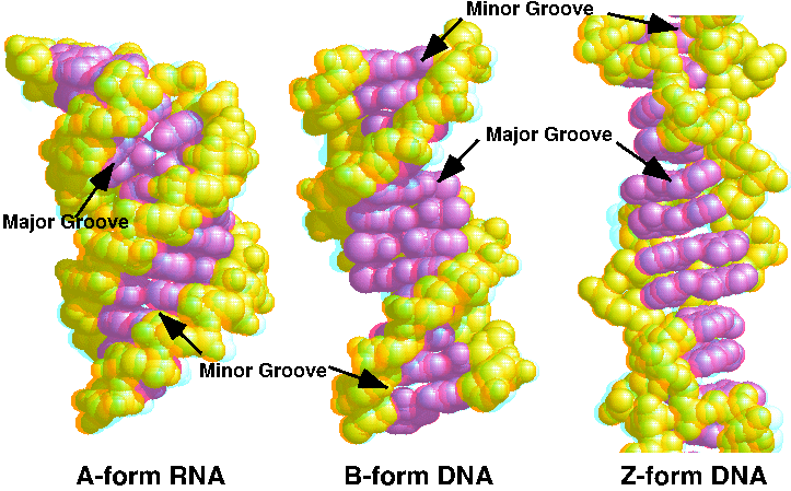
<LI> Narrow minor groove | <LI> Narrow minor groove | ||
<LI> Conformation favored by high salt concentrations, some base substitutions, but requires alternating purine-pyrimidine sequence. | <LI> Conformation favored by high salt concentrations, some base substitutions, but requires alternating purine-pyrimidine sequence. | ||
| - | <!--<LI> N2-amino of G H-bonds to 5' PO: explains slow exchange of proton, need for G purine. --> | ||
<LI> Base pairs nearly perpendicular to helix axis | <LI> Base pairs nearly perpendicular to helix axis | ||
<LI> GpC repeat, not single base-pair | <LI> GpC repeat, not single base-pair | ||
| Line 20: | Line 19: | ||
<LI> Conformations: | <LI> Conformations: | ||
<UL> | <UL> | ||
| - | <LI>G; syn, C2'-endo | + | <LI>G; ''syn'', C2'-''endo'' |
| - | <LI>C; anti, C3'-endo | + | <LI>C; ''anti'', C3'-''endo'' |
</UL> | </UL> | ||
</UL> | </UL> | ||
| Line 27: | Line 26: | ||
== Take the Tour == | == Take the Tour == | ||
| - | The tour starts with the <scene name='72/ | + | The tour starts with the <scene name='72/725870/Z-dna_overview/1'>Default</scene> view. Now look at this <scene name='72/725870/Space_filling_view/1'>space filling view</scene>.The backbone is yellow and the bases are magenta. Note that the major groove (at the top, when you have just clicked the button) is so wide that it is not really a groove any more. |
| - | Now change the display to make it show the <scene name='72/ | + | Now change the display to make it show the <scene name='72/725870/Space_filling_bbone/1'>sugar-phosphate backbone as pseudo-bonds</scene> connecting the phosphate atoms. Now the bases are easier to see. Now the bases are easier to see. Notice how they are stacked upon each other and are nearly perpendicular to the axis of the double helix. But notice that the base pairs do not stack upon each other equivalently. The backbone also is not a continuous curve, it "zig-zags" back and forth (hence "Z"-DNA). |
You can <scene name='72/725442/Zoom_pairs/1'>look at just four of the base pairs.</scene>.You are looking into the major groove and the colors of the base pairs alternate. You can also <scene name='72/725442/Zoom_pairs_only/1'>looks at just the bases</scene>. | You can <scene name='72/725442/Zoom_pairs/1'>look at just four of the base pairs.</scene>.You are looking into the major groove and the colors of the base pairs alternate. You can also <scene name='72/725442/Zoom_pairs_only/1'>looks at just the bases</scene>. | ||
Revision as of 20:16, 21 February 2016
Z-form DNA model
| |||||||||||
References
R. E. Dickerson, H. R. Drew, B. N. Conner, R. M. Wing, A. V. Fratini & M. L. Kopka (1982) The anatomy of A-, B-, and Z-DNA. Science 216: 475-485 [1] JSmol in Proteopedia [2] or to the article describing Jmol [3] to the rescue.
