This old version of Proteopedia is provided for student assignments while the new version is undergoing repairs. Content and edits done in this old version of Proteopedia after March 1, 2026 will eventually be lost when it is retired in about June of 2026.
Apply for new accounts at the new Proteopedia. Your logins will work in both the old and new versions.
B-DNA tour
From Proteopedia
(Difference between revisions)
| Line 1: | Line 1: | ||
==B-form DNA== | ==B-form DNA== | ||
<StructureSection load='1bna' size='400' side='right' caption='B-DNA' scene='72/725442/B-dna_overview/2'> | <StructureSection load='1bna' size='400' side='right' caption='B-DNA' scene='72/725442/B-dna_overview/2'> | ||
| + | Source <ref>PMID:7071593</ref> | ||
== Structural highlights == | == Structural highlights == | ||
<UL> | <UL> | ||
| Line 23: | Line 24: | ||
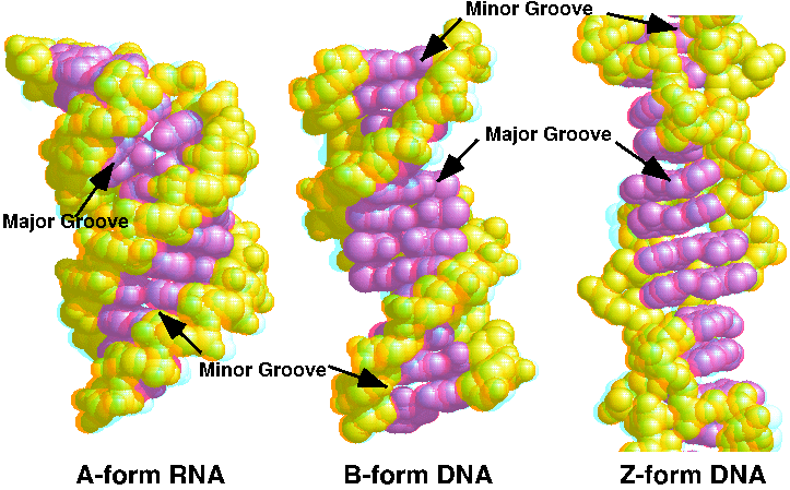
DNA is usually found in the B form under physiological conditions. The B-form conformation is stabilized by water molecules bound to the minor groove. You can see them as red dots <scene name='72/725442/Water_spine/1'>in this view</scene>. Sometimes kinks are found in the B helix at transcriptional control regions. These kinks can either be intrinsic to the DNA sequence or caused by transcription factor binding. | DNA is usually found in the B form under physiological conditions. The B-form conformation is stabilized by water molecules bound to the minor groove. You can see them as red dots <scene name='72/725442/Water_spine/1'>in this view</scene>. Sometimes kinks are found in the B helix at transcriptional control regions. These kinks can either be intrinsic to the DNA sequence or caused by transcription factor binding. | ||
| - | You can compare it with the DNA forms by looking at this [http://proteopedia.org/wiki/images/d/d3/JnABZ3d.gif 3D red-blue | + | You can compare it with the DNA forms by looking at this [http://proteopedia.org/wiki/images/d/d3/JnABZ3d.gif 3D red-blue stereo picture of A, B, and Z DNA] |
</StructureSection> | </StructureSection> | ||
==See Also== | ==See Also== | ||
| Line 32: | Line 33: | ||
* An interactive tutorial on [http://dna.molviz.org DNA Structure], ''disponible también en español'' and eight other languages. | * An interactive tutorial on [http://dna.molviz.org DNA Structure], ''disponible también en español'' and eight other languages. | ||
== References == | == References == | ||
| - | + | ||
| - | + | ||
JSmol in Proteopedia <ref>DOI 10.1002/ijch.201300024</ref> or to the article describing Jmol <ref>PMID:21638687</ref> to the rescue. | JSmol in Proteopedia <ref>DOI 10.1002/ijch.201300024</ref> or to the article describing Jmol <ref>PMID:21638687</ref> to the rescue. | ||
<references /> | <references /> | ||
Revision as of 13:42, 22 February 2016
B-form DNA
| |||||||||||
See Also
- Z-DNA model tour and Z-DNA
- A-RNA tour
- A more general overview will be found at DNA.
- Forms of DNA shows a side-by-side comparison of A, B, and Z forms of DNA.
- An interactive tutorial on DNA Structure, disponible también en español and eight other languages.
References
JSmol in Proteopedia [2] or to the article describing Jmol [3] to the rescue.
- ↑ Dickerson RE, Drew HR, Conner BN, Wing RM, Fratini AV, Kopka ML. The anatomy of A-, B-, and Z-DNA. Science. 1982 Apr 30;216(4545):475-85. PMID:7071593
- ↑ Hanson, R. M., Prilusky, J., Renjian, Z., Nakane, T. and Sussman, J. L. (2013), JSmol and the Next-Generation Web-Based Representation of 3D Molecular Structure as Applied to Proteopedia. Isr. J. Chem., 53:207-216. doi:http://dx.doi.org/10.1002/ijch.201300024
- ↑ Herraez A. Biomolecules in the computer: Jmol to the rescue. Biochem Mol Biol Educ. 2006 Jul;34(4):255-61. doi: 10.1002/bmb.2006.494034042644. PMID:21638687 doi:10.1002/bmb.2006.494034042644
