We apologize for Proteopedia being slow to respond. For the past two years, a new implementation of Proteopedia has been being built. Soon, it will replace this 18-year old system. All existing content will be moved to the new system at a date that will be announced here.
User:Daniel Schemenauer/Sandbox 1
From Proteopedia
(Difference between revisions)
| Line 18: | Line 18: | ||
===Interactions with a Negative Allosteric Modulator=== | ===Interactions with a Negative Allosteric Modulator=== | ||
The structure of mGlu<sub>5</sub> bound to the NAM [https://en.wikipedia.org/wiki/Mavoglurant Mavoglurant] demonstrates how protein activity is decreased through drug interactions. | The structure of mGlu<sub>5</sub> bound to the NAM [https://en.wikipedia.org/wiki/Mavoglurant Mavoglurant] demonstrates how protein activity is decreased through drug interactions. | ||
| - | <scene name='72/726404/Scene_7/2'>Mavoglurant</scene> binds within the allosteric binding site in the core of the seven trans-membrane α-helices,having passed through the restricted entrance formed by the ECL2. Bound Mavoglurant forms multiple interactions with the protein that further stabilize the inactive conformation. | + | <scene name='72/726404/Scene_7/2'>Mavoglurant</scene> binds within the allosteric binding site in the core of the seven trans-membrane α-helices,having passed through the restricted entrance formed by the <scene name='72/726409/Mavoglurant_overview2/3'>ECL2</scene>. Bound Mavoglurant forms multiple interactions with the protein that further stabilize the inactive conformation. |
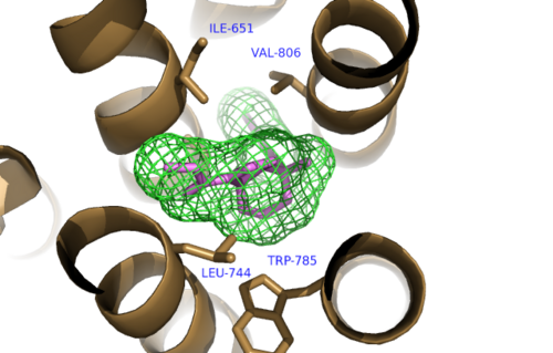
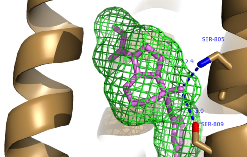
The bicyclic ring system of the drug is surrounded by a pocket of mainly hydrophobic residues including Val 806, Met 802, Phe 788, Trp 785, Leu 744, Ile 651, Pro 655 and Asn 747<ref name="Primary">PMID: 25042998 </ref> (Figure 1). The carbamate tail of mavoglurant forms a hydrogen bond through its carbonyl oxygen to the amide side-chain of Asparagine 747 of TM4 (Figure 2). A hydroxyl group similarly forms hydrogen bonds to the protein, specifically to two serine residues (S805 and S809) of TM7 which form a hydrogen bonding network to other residues through their main chain atoms and a coordinated water molecule (omitted for clarity) (Figure 3). The interactions between Mavoglurant andmGlu<sub>5</sub> involved TM helices that were not previously stabilized by any strong interactions, introducing a new level of stability that favors the inactive conformation of the protein and hence decrease activity<ref name="Primary">PMID: 25042998 </ref>. | The bicyclic ring system of the drug is surrounded by a pocket of mainly hydrophobic residues including Val 806, Met 802, Phe 788, Trp 785, Leu 744, Ile 651, Pro 655 and Asn 747<ref name="Primary">PMID: 25042998 </ref> (Figure 1). The carbamate tail of mavoglurant forms a hydrogen bond through its carbonyl oxygen to the amide side-chain of Asparagine 747 of TM4 (Figure 2). A hydroxyl group similarly forms hydrogen bonds to the protein, specifically to two serine residues (S805 and S809) of TM7 which form a hydrogen bonding network to other residues through their main chain atoms and a coordinated water molecule (omitted for clarity) (Figure 3). The interactions between Mavoglurant andmGlu<sub>5</sub> involved TM helices that were not previously stabilized by any strong interactions, introducing a new level of stability that favors the inactive conformation of the protein and hence decrease activity<ref name="Primary">PMID: 25042998 </ref>. | ||
[[Image:Mav_Hydrophobic_pocket.png |500 px|left|thumb|Figure 1.Hydrophobic Pocket Surrounding Mavoglurant]] | [[Image:Mav_Hydrophobic_pocket.png |500 px|left|thumb|Figure 1.Hydrophobic Pocket Surrounding Mavoglurant]] | ||
Revision as of 12:11, 29 March 2016
| |||||||||||
References
- ↑ Vassilatis DK, Hohmann JG, Zeng H, Li F, Ranchalis JE, Mortrud MT, Brown A, Rodriguez SS, Weller JR, Wright AC, Bergmann JE, Gaitanaris GA. The G protein-coupled receptor repertoires of human and mouse. Proc Natl Acad Sci U S A. 2003 Apr 15;100(8):4903-8. Epub 2003 Apr 4. PMID:12679517 doi:http://dx.doi.org/10.1073/pnas.0230374100
- ↑ 2.0 2.1 Venkatakrishnan AJ, Deupi X, Lebon G, Tate CG, Schertler GF, Babu MM. Molecular signatures of G-protein-coupled receptors. Nature. 2013 Feb 14;494(7436):185-94. doi: 10.1038/nature11896. PMID:23407534 doi:http://dx.doi.org/10.1038/nature11896
- ↑ Pin JP, Galvez T, Prezeau L. Evolution, structure, and activation mechanism of family 3/C G-protein-coupled receptors. Pharmacol Ther. 2003 Jun;98(3):325-54. PMID:12782243
- ↑ 4.0 4.1 4.2 4.3 4.4 4.5 4.6 4.7 4.8 4.9 Dore AS, Okrasa K, Patel JC, Serrano-Vega M, Bennett K, Cooke RM, Errey JC, Jazayeri A, Khan S, Tehan B, Weir M, Wiggin GR, Marshall FH. Structure of class C GPCR metabotropic glutamate receptor 5 transmembrane domain. Nature. 2014 Jul 31;511(7511):557-62. doi: 10.1038/nature13396. Epub 2014 Jul 6. PMID:25042998 doi:http://dx.doi.org/10.1038/nature13396
- ↑ Shigemoto R, Nomura S, Ohishi H, Sugihara H, Nakanishi S, Mizuno N. Immunohistochemical localization of a metabotropic glutamate receptor, mGluR5, in the rat brain. Neurosci Lett. 1993 Nov 26;163(1):53-7. PMID:8295733
- ↑ Li G, Jorgensen M, Campbell BM. Metabotropic glutamate receptor 5-negative allosteric modulators for the treatment of psychiatric and neurological disorders (2009-July 2013). Pharm Pat Anal. 2013 Nov;2(6):767-802. doi: 10.4155/ppa.13.58. PMID:24237242 doi:http://dx.doi.org/10.4155/ppa.13.58


