We apologize for Proteopedia being slow to respond. For the past two years, a new implementation of Proteopedia has been being built. Soon, it will replace this 18-year old system. All existing content will be moved to the new system at a date that will be announced here.
Sandbox Reserved 1160
From Proteopedia
(Difference between revisions)
| Line 8: | Line 8: | ||
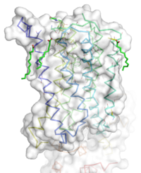
[[Image:STR.png|200 px|left|thumb|Overall Structure of the TMD. The polar heads on the Oliec acids orient the protein with the top of the image being the extracellular portion of the protein,the middle portion inserted into the membrane, and the lower portion located inside of the cell. ]] | [[Image:STR.png|200 px|left|thumb|Overall Structure of the TMD. The polar heads on the Oliec acids orient the protein with the top of the image being the extracellular portion of the protein,the middle portion inserted into the membrane, and the lower portion located inside of the cell. ]] | ||
=== Overview === | === Overview === | ||
| - | The mGlu5 TMD contains 7 <scene name='72/721531/Protien_7_helices/2'> alpha helices</scene> that span the membrane. The protein was crystallized with Oleic acid and MES. On the superior portion of the protein there are several critical extracellular loops.The binding pocket can be found near the middle of the protein.Inserted into the biding pocket is the negative allosteric modulator Mavoglurant. It is important to note that the TMD as illustrated is in an inactive conformation. On the intracellular portion of the protein there exist several ionic locks whose positions will determine the activity of the protein. | + | The mGlu5 TMD contains 7 <scene name='72/721531/Protien_7_helices/2'> alpha helices</scene> that span the membrane. The protein was crystallized with Oleic acid and MES. On the superior portion of the protein there are several critical extracellular loops.The binding pocket can be found near the middle of the protein.Inserted into the biding pocket is the negative allosteric modulator [http://www.en.wikipedia.org/wiki/Mavoglurant mavoglurant]. It is important to note that the TMD as illustrated is in an inactive conformation. On the intracellular portion of the protein there exist several ionic locks whose positions will determine the activity of the protein. |
=== Extracellular Domain === | === Extracellular Domain === | ||
=== Binding Pocket === | === Binding Pocket === | ||
| Line 18: | Line 18: | ||
*A <scene name='72/721531/Protien_bindbottom/1'>water molecular</scene> inside of the binding pocket helps stabilize the inactive state. | *A <scene name='72/721531/Protien_bindbottom/1'>water molecular</scene> inside of the binding pocket helps stabilize the inactive state. | ||
| - | Once bound to | + | Once bound to mavoglurant, transmembrane helix 7 undergoes a conformational change. The shifting of TM7 will lead to a more global conformational change, which we leave the receptor incapable of signaling. |
=== Ionic Locks === | === Ionic Locks === | ||
| - | Another important structural feature of the protein is the series of <scene name='72/721531/Ionic_lock/2'>ionic locks</scene> on the intracellular side of the protein. Interaction between amino acids will form a salt bridge which will stabilize the inactive conformation. The primary ionic lock forms between Glu770, Lys665, and Ser613. A secondary ionic lock occurs between Ser614 and Arg668. The purpose of these ionic locks are analogous to the ionic interactions that stabilize the T state in | + | Another important structural feature of the protein is the series of <scene name='72/721531/Ionic_lock/2'>ionic locks</scene> on the intracellular side of the protein. Interaction between amino acids will form a salt bridge which will stabilize the inactive conformation. The primary ionic lock forms between Glu770, Lys665, and Ser613. A secondary ionic lock occurs between Ser614 and Arg668. The purpose of these ionic locks are analogous to the ionic interactions that stabilize the T state in [[Hemoglobin]]. In the case of the TMD, when the NAM mavoglurant is bound the ionic lock is formed. This stabilizes the inactive state, where the intracellular loops are stabilized inwards. This will effectively block the crevice that is involved in binding the G-protien. Models have suggested that, even in a glutamate bound state, the mavoglurant bound receptor would be dimerized but incapable of signaling. This helps maintain the readiness of the pathway, while still decreasing signal response. |
== Function and Pathway == | == Function and Pathway == | ||
| Line 46: | Line 46: | ||
<references/> | <references/> | ||
== External Resources == | == External Resources == | ||
| + | [http://www.fraxa.org/novartis-discontinues-development-mavoglurant-afq056-fragile-x-syndrome/ Novartis Fragile X trials] | ||
Revision as of 02:34, 30 March 2016
Human metabotropic glutamate receptor 5 transmembrane domain
| |||||||||||
References
- ↑ 1.0 1.1 Dore AS, Okrasa K, Patel JC, Serrano-Vega M, Bennett K, Cooke RM, Errey JC, Jazayeri A, Khan S, Tehan B, Weir M, Wiggin GR, Marshall FH. Structure of class C GPCR metabotropic glutamate receptor 5 transmembrane domain. Nature. 2014 Jul 31;511(7511):557-62. doi: 10.1038/nature13396. Epub 2014 Jul 6. PMID:25042998 doi:http://dx.doi.org/10.1038/nature13396
- ↑ 2.0 2.1 2.2 Niswender CM, Conn PJ. Metabotropic glutamate receptors: physiology, pharmacology, and disease. Annu Rev Pharmacol Toxicol. 2010;50:295-322. doi:, 10.1146/annurev.pharmtox.011008.145533. PMID:20055706 doi:http://dx.doi.org/10.1146/annurev.pharmtox.011008.145533
- ↑ 3.0 3.1 3.2 Wu H, Wang C, Gregory KJ, Han GW, Cho HP, Xia Y, Niswender CM, Katritch V, Meiler J, Cherezov V, Conn PJ, Stevens RC. Structure of a class C GPCR metabotropic glutamate receptor 1 bound to an allosteric modulator. Science. 2014 Apr 4;344(6179):58-64. doi: 10.1126/science.1249489. Epub 2014 Mar , 6. PMID:24603153 doi:http://dx.doi.org/10.1126/science.1249489
- ↑ 4.0 4.1 4.2 4.3 4.4 Bailey DB Jr, Berry-Kravis E, Wheeler A, Raspa M, Merrien F, Ricart J, Koumaras B, Rosenkranz G, Tomlinson M, von Raison F, Apostol G. Mavoglurant in adolescents with fragile X syndrome: analysis of Clinical Global Impression-Improvement source data from a double-blind therapeutic study followed by an open-label, long-term extension study. J Neurodev Disord. 2016;8:1. doi: 10.1186/s11689-015-9134-5. Epub 2015 Dec 15. PMID:26855682 doi:http://dx.doi.org/10.1186/s11689-015-9134-5
- ↑ 5.0 5.1 Feng Z, Ma S, Hu G, Xie XQ. Allosteric Binding Site and Activation Mechanism of Class C G-Protein Coupled Receptors: Metabotropic Glutamate Receptor Family. AAPS J. 2015 May;17(3):737-53. doi: 10.1208/s12248-015-9742-8. Epub 2015 Mar 12. PMID:25762450 doi:http://dx.doi.org/10.1208/s12248-015-9742-8

