We apologize for Proteopedia being slow to respond. For the past two years, a new implementation of Proteopedia has been being built. Soon, it will replace this 18-year old system. All existing content will be moved to the new system at a date that will be announced here.
Sandbox Reserved 1160
From Proteopedia
(Difference between revisions)
| Line 10: | Line 10: | ||
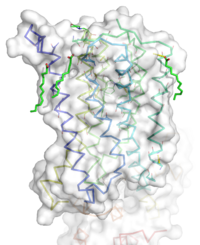
The mGlu5 TMD contains 7 <scene name='72/721531/Protien_7_helices/2'> alpha helices</scene> that span the membrane. The protein was crystallized with Oleic acid and MES. On the superior portion of the protein there are several critical extracellular loops.The binding pocket can be found near the middle of the protein.Inserted into the biding pocket is the negative allosteric modulator [http://www.en.wikipedia.org/wiki/Mavoglurant mavoglurant]. It is important to note that the TMD as illustrated is in an inactive conformation. On the intracellular portion of the protein there exist several ionic locks whose positions will determine the activity of the protein. | The mGlu5 TMD contains 7 <scene name='72/721531/Protien_7_helices/2'> alpha helices</scene> that span the membrane. The protein was crystallized with Oleic acid and MES. On the superior portion of the protein there are several critical extracellular loops.The binding pocket can be found near the middle of the protein.Inserted into the biding pocket is the negative allosteric modulator [http://www.en.wikipedia.org/wiki/Mavoglurant mavoglurant]. It is important to note that the TMD as illustrated is in an inactive conformation. On the intracellular portion of the protein there exist several ionic locks whose positions will determine the activity of the protein. | ||
=== Extracellular Domain === | === Extracellular Domain === | ||
| + | This is the <scene name='72/721532/Ecl_trail_1/7'>Extracellular Loops</scene> shows the extracellular loops (ECL) 1, 2, and 3 highlighted in purple. Additionally in the ECL domain, a <scene name='72/721532/Ecl_trail_1/6'>Disulfide Bond</scene> is attached to Helix 3 and the Amino Acid chain between Helix 5 and the N terminus. The disulfide bond is highlighted in yellow, and it is conserved in all classes of glutamate receptor 5 transmembrane domains. | ||
=== Binding Pocket === | === Binding Pocket === | ||
| - | The binding pocket represents an interesting source of regulatory control of receptor activity. The binding pocket is only accessible by a relatively narrow (7 angstrom) <scene name='72/721531/Protien_sur/4'>entrance</scene><ref name="Dore" />. This small entrance severely restricts the access of both positive and negative allosteric regulators. This structural feature will severely limit the size of possible regulators. | + | [[Image: Organic with clipped surface.png|100 px|left|thumb|Cross section view of mavoglurant in the binding pocket]] |
| + | The binding pocket represents an interesting source of regulatory control of receptor activity. The binding pocket is only accessible by a relatively narrow (7 angstrom) <scene name='72/721531/Protien_sur/4'>entrance</scene><ref name="Dore" />. This small entrance severely restricts the access of both positive and negative allosteric regulators. This structural feature will severely limit the size of possible regulators. Variation can be seen in positioning of alpha helices. Class C receptors has seemingly less space for [https://fragilex.org/2014/research/news-reports-and-commentaries/novartis-announces-results-of-mavoglurant-mglur5-afq056-clinical-trials-and-the-conclusion-of-the-long-term-extension-study/ mavoglurant] to enter compared to Class A and F . The ligand binding site is also varied between different classes of receptors. | ||
Important Amino Acids<ref name="Dore" />: | Important Amino Acids<ref name="Dore" />: | ||
*<scene name='72/721531/Protien_bindtop/4'>Asparagine</scene> 747forms a hydrogen bond network with the main chain carbonyl of Glycine 652 and the carbamate portion of mavoglurant. | *<scene name='72/721531/Protien_bindtop/4'>Asparagine</scene> 747forms a hydrogen bond network with the main chain carbonyl of Glycine 652 and the carbamate portion of mavoglurant. | ||
| Line 24: | Line 26: | ||
== Function and Pathway == | == Function and Pathway == | ||
| - | + | It all begins with glutamate binding to the venus fly trap domain. The signal transduction goes across the cystine-rich domain to the TMD. Next the dimerization of the TMD occurs. This activates the Gq/11 pathway, which activates phspholipase Cβ. The active phospholipase Cβ performs hydrolysis on phosphotinositides and generates inositol 1,4,5-trisphosphate and diacyl-glycerol. This results in calcium mobilization and activation of protein kinase C. | |
== Comparison to Similar Structures == | == Comparison to Similar Structures == | ||
== Disease == | == Disease == | ||
Revision as of 10:50, 30 March 2016
Human metabotropic glutamate receptor 5 transmembrane domain
| |||||||||||


