User:Dean Williams/Sandbox 1180
From Proteopedia
| Line 37: | Line 37: | ||
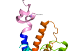
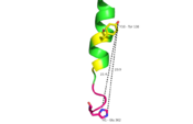
| - | Secondly, the extracellular loop 1 (ECL1) is 3-4 times longer than comparable loops in class A GPCRs, and also affects ligand binding affinity. (Fig. 3)<ref name= "Siu 2013"/> [[Image:Asn_298__Trp_295.png|150 px|left|thumb|Fig. 3: Active sites linked to glucagon binding affinity located on ECL1 are labeled<ref name= "Siu 2013"/>.]] | + | Secondly, the extracellular loop 1 (ECL1) is 3-4 times longer than comparable loops in class A GPCRs, and also affects ligand binding affinity. (Fig. 3)<ref name= "Siu 2013"/> [[Image:Asn_298__Trp_295.png|150 px|left|thumb|Fig. 3: Active sites linked to glucagon binding affinity located on ECL1 are labeled<ref name= "Siu 2013"/>.]] |
| Line 56: | Line 56: | ||
[[Image:Corticotropin class B and Glucagon class B receptors aligned.png |150 px|left|thumb|Fig. 4: Corticotropin-releasing factor 1 and glucagon receptors; Class B GPCRs with notable central splay]] [[Image:Beta2 class A and Glucagon class B receptors aligned.png |150 px|right|thumb|Fig. 5: Beta 2-adrenergic (class A) and glucagon receptors; showing an absence of central splay in Class A GPCRs.]] | [[Image:Corticotropin class B and Glucagon class B receptors aligned.png |150 px|left|thumb|Fig. 4: Corticotropin-releasing factor 1 and glucagon receptors; Class B GPCRs with notable central splay]] [[Image:Beta2 class A and Glucagon class B receptors aligned.png |150 px|right|thumb|Fig. 5: Beta 2-adrenergic (class A) and glucagon receptors; showing an absence of central splay in Class A GPCRs.]] | ||
| + | |||
| + | |||
| + | |||
| + | |||
| + | |||
| + | |||
| Line 88: | Line 94: | ||
===Peptide binding and selectivity=== | ===Peptide binding and selectivity=== | ||
It has been discovered that the large, soluble N-terminal extracellular domains (ECD) of GCGR are primary in ligand selectivity with the deep, ligand pocket (Fig. 7) of the TMD providing secondary recognition. <ref name= "Yang 2015"/> [[Image:Movie_Frame_2.png|100 px|left|thumb|Fig. 7: Active site buried deep in 7TMD of glucagon receptor.]] | It has been discovered that the large, soluble N-terminal extracellular domains (ECD) of GCGR are primary in ligand selectivity with the deep, ligand pocket (Fig. 7) of the TMD providing secondary recognition. <ref name= "Yang 2015"/> [[Image:Movie_Frame_2.png|100 px|left|thumb|Fig. 7: Active site buried deep in 7TMD of glucagon receptor.]] | ||
| + | |||
| + | |||
| + | |||
| + | |||
| + | |||
| + | |||
| + | |||
| + | |||
===Conformational changes=== | ===Conformational changes=== | ||
| Line 126: | Line 140: | ||
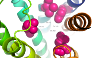
Four essential residues exist deep within the central cavity which all play strong roles in ligand binding affinity. (Fig. 9) | Four essential residues exist deep within the central cavity which all play strong roles in ligand binding affinity. (Fig. 9) | ||
[[Image:Movie Frame 3.png |200 px|right|thumb|Fig. 9: Location of anchoring pocket within central cavity.<ref name= "Siu 2013"/>]] | [[Image:Movie Frame 3.png |200 px|right|thumb|Fig. 9: Location of anchoring pocket within central cavity.<ref name= "Siu 2013"/>]] | ||
| + | |||
| + | |||
| + | |||
| + | |||
| + | |||
| + | |||
| + | |||
| + | |||
| + | |||
| + | |||
| + | |||
| + | |||
| + | |||
| + | |||
| + | |||
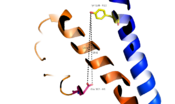
A narrow entry gives way to a large, anchoring site for residues 1-4 of glucagon. (Fig. 10) | A narrow entry gives way to a large, anchoring site for residues 1-4 of glucagon. (Fig. 10) | ||
[[Image:Movie Frame 6.png |100 px|left|thumb|Fig. 10: Ballooned pocket functioning as anchoring site for glucagon residues 1-4.]] | [[Image:Movie Frame 6.png |100 px|left|thumb|Fig. 10: Ballooned pocket functioning as anchoring site for glucagon residues 1-4.]] | ||
| - | Essential to glucagon's | + | Essential to glucagon's binding, a long, N-terminal tail winds to a clump of 4 residues, culminating in bulge that fits into the central, anchoring site of the 7TMD. (Fig. 11) |
[[Image:Glucagon with Q3 and N-terminus.png |200 px|right|thumb|Fig. 11: Surface visualization of glucagon demonstrating three dimensional fit of N-terminal tail into binding site of GCGR central cavity active site]] | [[Image:Glucagon with Q3 and N-terminus.png |200 px|right|thumb|Fig. 11: Surface visualization of glucagon demonstrating three dimensional fit of N-terminal tail into binding site of GCGR central cavity active site]] | ||
| + | |||
| + | |||
| + | |||
| + | |||
| + | |||
| + | |||
| + | |||
| + | |||
| Line 153: | Line 190: | ||
[[Image:Small molecule modulators Page 1.jpg|150 px|left|thumb|Fig. 12: Small molecule regulators of GCGR, part 1<ref name= "Yang 2015"/>.]] | [[Image:Small molecule modulators Page 1.jpg|150 px|left|thumb|Fig. 12: Small molecule regulators of GCGR, part 1<ref name= "Yang 2015"/>.]] | ||
| - | [[Image:Small molecule modulators Page 2.jpg|150 px|right|thumb|Fig. 13: Small molecule regulators of GCGR, part | + | [[Image:Small molecule modulators Page 2.jpg|150 px|right|thumb|Fig. 13: Small molecule regulators of GCGR, part 2<ref name= "Yang 2015"/>.]] |
| + | |||
| + | |||
| + | |||
| + | |||
| + | |||
| + | |||
| + | |||
| + | |||
| + | |||
| + | |||
| + | |||
| + | |||
| + | |||
| + | |||
| + | |||
| + | |||
| + | |||
| + | |||
| + | |||
| + | |||
| + | |||
===Possible structural considerations for large molecule agonists/antagonists=== | ===Possible structural considerations for large molecule agonists/antagonists=== | ||
Revision as of 21:11, 2 April 2016
Structure of Class B Human Glucagon G-Protein Coupled Receptors (GCGRs)
G protein coupled receptors (GPCRs) are recognized as the largest known class of integral membrane proteins and are divided into five families; the rhodopsin family (class A), the secretin family (class B), the adhesion family, the glutamate family (class C), and the frizzled/taste family (class F). Roughly 5% of the human genome encodes g-protein-coupled receptors which are responsible for the transduction of endogenous signals and the instigation of cellular response. The variants all contain a similar seven α-helical transmembrane domain (TMD or 7TMD) that, once bound to its peptide ligand, undergoes conformational change and tranduces a signal to coupled, heterotrimeric G proteins which initiate intracellular signal pathways and generate physiological and pathological processes. [1]
Class B GPCRs contain 15 distinct receptors for peptide hormones and generate their signal pathway through the activation of adenylate cyclase (AC) which increases concentration of cAMP, inositol phosphate, and calcium levels in cyto. [2] These signals are essential elements of intracellular signal cascades for human diseases including type II diabetes mellitus, osteoporosis, obesity, cancer, neurological degeneration, cardiovascular diseases, headaches, and psychiatric disorders; making their regulation through drug targeting of particular interest to companies developing novel molecules. [3] Structurally based approaches to the development of small-molecule agonists and antagonists have been hampered by the lack of accurate Class B TMD visualizations until recent crystal structures of corticoptropin-releasing factor receptor 1 and human glucagon were realized. [4] [5]
The glucagon class B GPCR (GCGR) is involved in glucose homeostasis through the binding of the signal peptide glucagon.
| |||||||||||
References
- ↑ Zhang Y, Devries ME, Skolnick J. Structure modeling of all identified G protein-coupled receptors in the human genome. PLoS Comput Biol. 2006 Feb;2(2):e13. Epub 2006 Feb 17. PMID:16485037 doi:http://dx.doi.org/10.1371/journal.pcbi.0020013
- ↑ Bortolato A, Dore AS, Hollenstein K, Tehan BG, Mason JS, Marshall FH. Structure of Class B GPCRs: new horizons for drug discovery. Br J Pharmacol. 2014 Jul;171(13):3132-45. doi: 10.1111/bph.12689. PMID:24628305 doi:http://dx.doi.org/10.1111/bph.12689
- ↑ 3.0 3.1 Hollenstein K, de Graaf C, Bortolato A, Wang MW, Marshall FH, Stevens RC. Insights into the structure of class B GPCRs. Trends Pharmacol Sci. 2014 Jan;35(1):12-22. doi: 10.1016/j.tips.2013.11.001. Epub, 2013 Dec 18. PMID:24359917 doi:http://dx.doi.org/10.1016/j.tips.2013.11.001
- ↑ Hollenstein K, Kean J, Bortolato A, Cheng RK, Dore AS, Jazayeri A, Cooke RM, Weir M, Marshall FH. Structure of class B GPCR corticotropin-releasing factor receptor 1. Nature. 2013 Jul 25;499(7459):438-43. doi: 10.1038/nature12357. Epub 2013 Jul 17. PMID:23863939 doi:http://dx.doi.org/10.1038/nature12357
- ↑ 5.0 5.1 5.2 5.3 5.4 5.5 5.6 5.7 Siu FY, He M, de Graaf C, Han GW, Yang D, Zhang Z, Zhou C, Xu Q, Wacker D, Joseph JS, Liu W, Lau J, Cherezov V, Katritch V, Wang MW, Stevens RC. Structure of the human glucagon class B G-protein-coupled receptor. Nature. 2013 Jul 25;499(7459):444-9. doi: 10.1038/nature12393. Epub 2013 Jul 17. PMID:23863937 doi:10.1038/nature12393
- ↑ 6.0 6.1 6.2 6.3 6.4 Yang DH, Zhou CH, Liu Q, Wang MW. Landmark studies on the glucagon subfamily of GPCRs: from small molecule modulators to a crystal structure. Acta Pharmacol Sin. 2015 Sep;36(9):1033-42. doi: 10.1038/aps.2015.78. Epub 2015, Aug 17. PMID:26279155 doi:http://dx.doi.org/10.1038/aps.2015.78
- ↑ Ahren B. Islet G protein-coupled receptors as potential targets for treatment of type 2 diabetes. Nat Rev Drug Discov. 2009 May;8(5):369-85. doi: 10.1038/nrd2782. Epub 2009 Apr, 14. PMID:19365392 doi:http://dx.doi.org/10.1038/nrd2782
- ↑ 8.0 8.1 Xu Y, Xie X. Glucagon receptor mediates calcium signaling by coupling to G alpha q/11 and G alpha i/o in HEK293 cells. J Recept Signal Transduct Res. 2009 Dec;29(6):318-25. doi:, 10.3109/10799890903295150. PMID:19903011 doi:http://dx.doi.org/10.3109/10799890903295150
- ↑ Weston C, Lu J, Li N, Barkan K, Richards GO, Roberts DJ, Skerry TM, Poyner D, Pardamwar M, Reynolds CA, Dowell SJ, Willars GB, Ladds G. Modulation of Glucagon Receptor Pharmacology by Receptor Activity-modifying Protein-2 (RAMP2). J Biol Chem. 2015 Sep 18;290(38):23009-22. doi: 10.1074/jbc.M114.624601. Epub, 2015 Jul 21. PMID:26198634 doi:http://dx.doi.org/10.1074/jbc.M114.624601
- ↑ Zhang X, Stevens RC, Xu F. The importance of ligands for G protein-coupled receptor stability. Trends Biochem Sci. 2015 Feb;40(2):79-87. doi: 10.1016/j.tibs.2014.12.005. Epub, 2015 Jan 15. PMID:25601764 doi:http://dx.doi.org/10.1016/j.tibs.2014.12.005
- ↑ 'Lehninger A., Nelson D.N, & Cox M.M. (2008) Lehninger Principles of Biochemistry. W. H. Freeman, fifth edition.'
- ↑ 12.0 12.1 Salon JA, Lodowski DT, Palczewski K. The significance of G protein-coupled receptor crystallography for drug discovery. Pharmacol Rev. 2011 Dec;63(4):901-37. doi: 10.1124/pr.110.003350. PMID:21969326 doi:http://dx.doi.org/10.1124/pr.110.003350
