This old version of Proteopedia is provided for student assignments while the new version is undergoing repairs. Content and edits done in this old version of Proteopedia after March 1, 2026 will eventually be lost when it is retired in about June of 2026.
Apply for new accounts at the new Proteopedia. Your logins will work in both the old and new versions.
LDL receptor
From Proteopedia
(Difference between revisions)
| Line 1: | Line 1: | ||
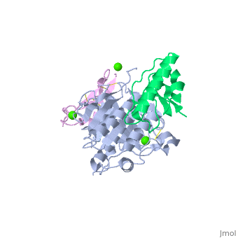
<StructureSection load='2w2m' size='350' side='right' caption='Structure of human LDLR EGF precursor homology domain (magenta) complex with proprotein convertase subtilisin/kexin 9-CoA reductase (grey and green) and Ca+2 ions (PDB entry [[2w2m]])' scene=''> | <StructureSection load='2w2m' size='350' side='right' caption='Structure of human LDLR EGF precursor homology domain (magenta) complex with proprotein convertase subtilisin/kexin 9-CoA reductase (grey and green) and Ca+2 ions (PDB entry [[2w2m]])' scene=''> | ||
| + | == Function == | ||
| + | '''LDL (Low Density Lipoprotein) receptor''' (LDLR) mediates the endocytosis of cholesterol-rich LDL. LDLR recognizes the apoprotein B100 which is embedded in the outer layer of the LDL particle. LDLR sits on the cell surface and binds LDL particles which circulate in the blood stream. LDLR transports the LDL particle into the cell where the cholesterol is used. Upon release of the LDL particle, the LDLR is recycled back into the cell membrane surface<ref>PMID:19299327</ref>. | ||
| - | + | == Structural highlights == | |
| + | LDLR consist of a ligand-binding domain (LBD residues 1-292), epidermal growth factor precursor homology domain (EGFP residues 293-699), oligosaccharide-rich domain (residues 700-758), membrane-spanning domain (residues 759-781) and cytoplasmic domain (residues 782-832). LDLR LBD contains 7 ca. 40 amino acid long repeats (LB) containing 6 cysteine residues, making a calcium binding octahedral structure. LDLR EGFP contains 2 EGF repeats followed by 6 YWTD repeats and another EGF repeat. LDLR LBD residues 133-273 are named C-type lectin-like domain. | ||
</StructureSection> | </StructureSection> | ||
| Line 53: | Line 56: | ||
**[[3p5b]], [[3p5c]] – hLDLR EGFP, oligosaccharide-rich, domains + proprotein convertase subtilisin/kexin 9<br /> | **[[3p5b]], [[3p5c]] – hLDLR EGFP, oligosaccharide-rich, domains + proprotein convertase subtilisin/kexin 9<br /> | ||
}} | }} | ||
| + | == References == | ||
| + | <references/> | ||
[[Category:Topic Page]] | [[Category:Topic Page]] | ||
Revision as of 09:54, 10 April 2016
| |||||||||||
3D structures of LDL receptor
Updated on 10-April-2016
References
- ↑ Goldstein JL, Brown MS. The LDL receptor. Arterioscler Thromb Vasc Biol. 2009 Apr;29(4):431-8. doi:, 10.1161/ATVBAHA.108.179564. PMID:19299327 doi:http://dx.doi.org/10.1161/ATVBAHA.108.179564
