Sandbox Reserved 1174
From Proteopedia
| Line 19: | Line 19: | ||
The LPA<sub>1</sub> receptor consists of seven transmembrane alpha helices. It lies in the membrane as shown in Figure 2, and as shown by the <scene name='72/721545/Membrane/4'>fatty acid</scene> bound in the crystallization of LPA<sub>1</sub> in orange. Most <scene name='72/721545/Polarity/3'>polar</scene> (red) reside on the intracellular and extracellular areas of the receptor, while most residues positioned on the trans membrane helices inside the membrane are hydrophobic (blue). | The LPA<sub>1</sub> receptor consists of seven transmembrane alpha helices. It lies in the membrane as shown in Figure 2, and as shown by the <scene name='72/721545/Membrane/4'>fatty acid</scene> bound in the crystallization of LPA<sub>1</sub> in orange. Most <scene name='72/721545/Polarity/3'>polar</scene> (red) reside on the intracellular and extracellular areas of the receptor, while most residues positioned on the trans membrane helices inside the membrane are hydrophobic (blue). | ||
| - | [[Image: | + | [[Image:LPA_in_membrane3.fw.png|200px|center|thumb|'''Figure 2:''' LPA in the Phospholipid Bilayer]] |
=== Structural Stabilization === | === Structural Stabilization === | ||
Revision as of 12:34, 12 April 2016
| This Sandbox is Reserved from Jan 11 through August 12, 2016 for use in the course CH462 Central Metabolism taught by R. Jeremy Johnson at the Butler University, Indianapolis, USA. This reservation includes Sandbox Reserved 1160 through Sandbox Reserved 1184. |
To get started:
More help: Help:Editing |
Contents |
Human Lysophosphatidic Acid Receptor 1
Lysophosphatidic Acid
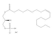
Lysophosphatidic Acid (LPA) consists of an unsaturated fatty acid chain, a glycerol backbone, and a free phosphate group (Figure 1). Lysophosphatidic acid is found in nearly all cells, tissues, and fluids of the body.[1] LPA is present intracellularly as a precursor of phospholipid biosynthesis, and extracellularly as a signalling phospholipid.
Extracellularly, LPA is produced from lysophosphatidylcholine by the enzyme autotaxin.[1] Autotaxin was originally linked with metastasis, and this link was later discovered to be mediated through the production of LPA, which signals cell proliferation.[2] All of LPA’s activities are receptor mediated; the signalling lipid interacts with at least six G-protein coupled receptors LPA1-LPA6.
Structure
| |||||||||||
Function
Of the six LPA G-protein coupled receptors, Lysophosphatidic acid recptor 1 (LPA1) is the most widely expressed.[4] LPA,1 is coupled to a heterotrimeric G protein on the intracellular side of the cell membrane. The three G alpha proteins that LPA1 couples to are Gi, Gq, and G12/13.[3] From these three G proteins many signal transduction pathways are activated. The downstream effects of Gi include cell proliferation, survival, and migration.[4] Gi leads to cell proliferation by the activation of the RAS-mediated MAPK cascade. [4] The alpha subunit Gq signals the inhibition of gap-junctional communication. The pathways activated by G12/13 include cell proliferation and morphology.[4] These downstream functions show the wide array of effects that LPA can have on the body. Targeted deletion of LPA receptors has had an effect on every organ system examined.[1]
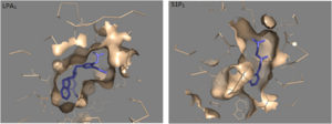
LPA1 is part of the larger EDG (endothelial differentiation gene) family, which includes the sphingosine 1-phosphate receptors. Significantly more research has been done on S1P1 than other receptors in this family. S1P1 is coupled to the same heterotrimeric G protein that LPA1 is, and both receptors are involved in growth-related activity and cytoskeletal functions. [5]
Clinical Relevance
Thus far the LPA receptors have had physiological effects on every organism that it has been tested with. There have been studies done looking at what happens with infertility, fibrosis, pain, and cancer when they come into contact with LPA receptors, and what happens when LPA receptors are deleted [1]. LPA receptors are commonly found in serum and saliva. [3].
Cancer
Many of the functions of LPA1, i.e. cell proliferation, survival, and morphology, are implicated in cancers. LPA has been shown to act as a tumor mitogen and an inducer of tumor-derived cytokine to support the metastasis (spreading) of breast and ovarian cancer to bones. Inhibition of LPA1 can significantly reduce this progression, and therefore may be a promising treatment for patients with bone metastasis. [6] It has not been shown to have an effect on primary tumor size. [7]
Pain
When an injury occurs LPA is released in the body. It then will activate G-protein-coupled receptors. Within the nervous system, LPA plays a role in the nociceptive process (nociceptive pain is a sharp pain that can come from a mild burn or twisted ankle). The LPA signaling will activate GTPase RhoA. Once activated Rho translocates to the plasma membrane. Rho will activate Rho kinase (ROCK). The actiavtion of ROCK is a required step in the pathway in the stimulation of neurotic pain. When ROCK was inhibited it meant that the rest of the pathway would not work according to plan. Mice with the deletation of LPA1 receptors were studied to see the role that LPA signaling played in pain. In a study done with mice, those without the LPA1 receptor had lower levels of pain.[8]. Another use of LPA is it can help in stimulation of cell migration [3].
Fibrosis
To gain a better understand the role the LPA plays in fibrosis, a study was done with mice who had contracted fibrosis [9]. Idiopathic pulmonary fibrosis (IPF) has high rates of mortality. Research has been done to study the pathway of the LPA-LPA1 in fibroblast migration (Wound Healing). In the injured lungs, the IPF, fibroblast can be activated. In the lungs genes related to cell migration can be unregulated. The fibroblast migration can be regulated by LPA. The bronchoalveolar lavage (BAL) in mice that had fibrosis was elevated. The research supported the hypothesis that LPA1 plays an active role between lung injury and contracting pulmonary fibrosis. LPA has the ability to lead to a vascular leak after an initial injury which can lead to fibrosis. This study's findings shows that LPA1 is part of a link between lung injury and pulmonary fibrosis [9].
Endocannabinoids
The endocannabinoid system, located in the mammalian nervous system, regulates a variety of physiological processes including appetite, pain sensation, mood, and memory. Endocannabinoids, the natural ligands for cannabinoid receptors, are similar in structure to lysophosphatidic acid.[1] Both the cannabinoid receptors and the LPA receptors have a preference for long unsaturated acyl chains. The polar amino acid in the binding pocket is unique to the lysophospholipid and cannabinoid receptors.
A major cannabinoid signaling molecule, 2-arachidonyl glycerol (2-AG, Figure 4), can be phosphorylated into 2-arachidonyl phosphatidic acid (2-ALPA). 2-ALPA has a similar structure to LPA, and is able to bind in the LPA1 receptor binding pocket. 2-ALPA binding to LPA1 causes the same downstream signaling that the LPA molecule does, effectively connecting these two systems. Promiscuous ligand binding between these two pathways has potential functional and therapeutic implications.[1]
References
- ↑ 1.0 1.1 1.2 1.3 1.4 1.5 1.6 1.7 1.8 Chrencik JE, Roth CB, Terakado M, Kurata H, Omi R, Kihara Y, Warshaviak D, Nakade S, Asmar-Rovira G, Mileni M, Mizuno H, Griffith MT, Rodgers C, Han GW, Velasquez J, Chun J, Stevens RC, Hanson MA. Crystal Structure of Antagonist Bound Human Lysophosphatidic Acid Receptor 1. Cell. 2015 Jun 18;161(7):1633-43. doi: 10.1016/j.cell.2015.06.002. PMID:26091040 doi:http://dx.doi.org/10.1016/j.cell.2015.06.002
- ↑ Boutin JA, Ferry G. Autotaxin. Cell Mol Life Sci. 2009 Sep;66(18):3009-21. doi: 10.1007/s00018-009-0056-9. Epub , 2009 Jun 9. PMID:19506801 doi:http://dx.doi.org/10.1007/s00018-009-0056-9
- ↑ 3.0 3.1 3.2 3.3 Moolenaar WH, van Meeteren LA, Giepmans BN. The ins and outs of lysophosphatidic acid signaling. Bioessays. 2004 Aug;26(8):870-81. PMID:15273989 doi:http://dx.doi.org/10.1002/bies.20081
- ↑ 4.0 4.1 4.2 4.3 Mills GB, Moolenaar WH. The emerging role of lysophosphatidic acid in cancer. Nat Rev Cancer. 2003 Aug;3(8):582-91. PMID:12894246 doi:http://dx.doi.org/10.1038/nrc1143
- ↑ Goetzl EJ, An S. Diversity of cellular receptors and functions for the lysophospholipid growth factors lysophosphatidic acid and sphingosine 1-phosphate. FASEB J. 1998 Dec;12(15):1589-98. PMID:9837849
- ↑ Boucharaba A, Serre CM, Guglielmi J, Bordet JC, Clezardin P, Peyruchaud O. The type 1 lysophosphatidic acid receptor is a target for therapy in bone metastases. Proc Natl Acad Sci U S A. 2006 Jun 20;103(25):9643-8. Epub 2006 Jun 12. PMID:16769891 doi:http://dx.doi.org/10.1073/pnas.0600979103
- ↑ Marshall JC, Collins JW, Nakayama J, Horak CE, Liewehr DJ, Steinberg SM, Albaugh M, Vidal-Vanaclocha F, Palmieri D, Barbier M, Murone M, Steeg PS. Effect of inhibition of the lysophosphatidic acid receptor 1 on metastasis and metastatic dormancy in breast cancer. J Natl Cancer Inst. 2012 Sep 5;104(17):1306-19. doi: 10.1093/jnci/djs319. Epub, 2012 Aug 21. PMID:22911670 doi:http://dx.doi.org/10.1093/jnci/djs319
- ↑ Inoue M, Rashid MH, Fujita R, Contos JJ, Chun J, Ueda H. Initiation of neuropathic pain requires lysophosphatidic acid receptor signaling. Nat Med. 2004 Jul;10(7):712-8. Epub 2004 Jun 13. PMID:15195086 doi:http://dx.doi.org/10.1038/nm1060
- ↑ 9.0 9.1 Tager AM, LaCamera P, Shea BS, Campanella GS, Selman M, Zhao Z, Polosukhin V, Wain J, Karimi-Shah BA, Kim ND, Hart WK, Pardo A, Blackwell TS, Xu Y, Chun J, Luster AD. The lysophosphatidic acid receptor LPA1 links pulmonary fibrosis to lung injury by mediating fibroblast recruitment and vascular leak. Nat Med. 2008 Jan;14(1):45-54. Epub 2007 Dec 9. PMID:18066075 doi:http://dx.doi.org/10.1038/nm1685


