We apologize for Proteopedia being slow to respond. For the past two years, a new implementation of Proteopedia has been being built. Soon, it will replace this 18-year old system. All existing content will be moved to the new system at a date that will be announced here.
Sandbox Reserved 1181
From Proteopedia
(Difference between revisions)
| Line 10: | Line 10: | ||
<scene name='72/721552/Glucagon_binding_zoomed_in/1'>Glucagon Binding Zoomed in</scene> | <scene name='72/721552/Glucagon_binding_zoomed_in/1'>Glucagon Binding Zoomed in</scene> | ||
| + | |||
| + | Old Stuff: | ||
| + | |||
| + | [[Image:ALA135PRO stalk unwinding.png|150 px|left|thumb|Fig. 1: A135P Mutation and effect on stalk stability <ref name= "Siu 2013"/>. ]] [[Image:Gln142 Tyr138 Glucagon interaction.png|150 px|right|thumb|Fig. 2: Stalk stabilized by salt bridge between Glu133-Lys136. Residues in yellow are demonstrated to have an effect on ligand binding affinity.<ref name= "Siu 2013"/>]] | ||
| + | |||
| + | [[Image:Asn_298__Trp_295.png|150 px|left|thumb|Fig. 3: Active sites linked to glucagon binding affinity located on ECL1 are labeled<ref name= "Siu 2013"/>.]] | ||
| + | |||
| + | [[Image:Corticotropin class B and Glucagon class B receptors aligned.png |150 px|left|thumb|Fig. 4: Corticotropin-releasing factor 1 and glucagon receptors; Class B GPCRs with notable central splay]] [[Image:Beta2 class A and Glucagon class B receptors aligned.png |150 px|right|thumb|Fig. 5: Beta 2-adrenergic (class A) and glucagon receptors; showing an absence of central splay in Class A GPCRs.]] | ||
| + | |||
| + | [[Image:Movie_Frame_2.png|100 px|left|thumb|Fig. 7: Active site buried deep in 7TMD of glucagon receptor.]] | ||
| + | |||
| + | [[Image:Movie Frame 3.png |200 px|right|thumb|Fig. 9: Location of anchoring pocket within central cavity.<ref name= "Siu 2013"/>]] | ||
| + | |||
| + | [[Image:Movie Frame 6.png |100 px|left|thumb|Fig. 10: Ballooned pocket functioning as anchoring site for glucagon residues 1-4.]] | ||
Revision as of 17:54, 20 April 2016
|
Old Stuff:

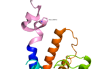
Fig. 1: A135P Mutation and effect on stalk stability [1].

Fig. 2: Stalk stabilized by salt bridge between Glu133-Lys136. Residues in yellow are demonstrated to have an effect on ligand binding affinity.[1]

Fig. 3: Active sites linked to glucagon binding affinity located on ECL1 are labeled[1].

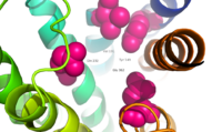
Fig. 9: Location of anchoring pocket within central cavity.[1]


