We apologize for Proteopedia being slow to respond. For the past two years, a new implementation of Proteopedia has been being built. Soon, it will replace this 18-year old system. All existing content will be moved to the new system at a date that will be announced here.
Sandbox Reserved 1181
From Proteopedia
(Difference between revisions)
| Line 1: | Line 1: | ||
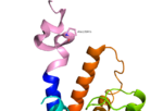
<Structure load='4L6R' size='350' frame='true' align='right' caption='7TM structure of human class B GPCR 4L6R' scene='72/727091/Full_structure_with_labels/1'/> | <Structure load='4L6R' size='350' frame='true' align='right' caption='7TM structure of human class B GPCR 4L6R' scene='72/727091/Full_structure_with_labels/1'/> | ||
| + | |||
| + | <scene name='72/727091/Full_Structure_with_Labels/1'>Labels</scene> | ||
<scene name='72/721552/Ligand_binding_interactions/1'>Ligand Binding Interactions and Crucial Disulfide Bond</scene> | <scene name='72/721552/Ligand_binding_interactions/1'>Ligand Binding Interactions and Crucial Disulfide Bond</scene> | ||
| - | |||
| - | <scene name='72/721552/Glucagon_binding/1'>Glucagon Binding Zoomed Out</scene> | ||
| - | |||
| - | <scene name='72/721552/Glucagon_binding/2'>Glucagon Binding Zoomed Out</scene> | ||
<scene name='72/721552/Glucagon_binding/3'>Glucagon Binding Full Rendering</scene> | <scene name='72/721552/Glucagon_binding/3'>Glucagon Binding Full Rendering</scene> | ||
| Line 20: | Line 18: | ||
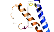
[[Image:Movie_Frame_2.png|100 px|left|thumb|Fig. 7: Active site buried deep in 7TMD of glucagon receptor.]] | [[Image:Movie_Frame_2.png|100 px|left|thumb|Fig. 7: Active site buried deep in 7TMD of glucagon receptor.]] | ||
| - | |||
| - | [[Image:Movie Frame 3.png |200 px|right|thumb|Fig. 9: Location of anchoring pocket within central cavity.<ref name= "Siu 2013"/>]] | ||
| - | |||
| - | [[Image:Movie Frame 6.png |100 px|left|thumb|Fig. 10: Ballooned pocket functioning as anchoring site for glucagon residues 1-4.]] | ||
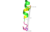
[[Image:Movie_Frame_7.png|175 px|left|thumb|Fig. 14: Distance measurement of GCGR 7TMD Y138-D362 of 19-20 angstroms and labeled with complimentary glucagon interaction residues.]] | [[Image:Movie_Frame_7.png|175 px|left|thumb|Fig. 14: Distance measurement of GCGR 7TMD Y138-D362 of 19-20 angstroms and labeled with complimentary glucagon interaction residues.]] | ||
Current revision
|
Old Stuff:

Fig. 1: A135P Mutation and effect on stalk stability [1].

Fig. 2: Stalk stabilized by salt bridge between Glu133-Lys136. Residues in yellow are demonstrated to have an effect on ligand binding affinity.[1]

Fig. 3: Active sites linked to glucagon binding affinity located on ECL1 are labeled[1].
Future research direction
Research for Class A GPCRs is much more extensive than for its secretin, class B counterparts, although class B is proving to be a worthwhile to invest researching. The challenge of class B stabilization, expression, and molecular size , has made class B GPCRs particularly hard to assay. Biochemical research has increased in the class B specifications, because it has been realized that receptors can be modulated by more than the agonist and antagonists present in vivo. Leading research consists of a complex interwoven scheme of equilibria manipulation in multi-receptor conformations. [2]


